An extramedullary positioning device for knee joint replacement femur

A knee joint replacement and positioning device technology, applied in joint implants, joint implants and other directions, can solve the problems of increasing the risk of surgical infection, prolonging the operation time, whether the positioning is rough, and achieving the convenience of large-scale promotion and use, The overall structure is ingeniously designed to reduce the pain of patients

Active Publication Date: 2016-08-24
THE AFFILIATED DRUM TOWER HOSPITAL MEDICAL SCHOOL OF NANJING UNIV
View PDF7 Cites 0 Cited by
  • Summary
  • Abstract
  • Description
  • Claims
  • Application Information

AI Technical Summary

Problems solved by technology

[0003] Traditional femoral intramedullary positioning increases intramedullary hemorrhage, and is closely related to the needle entry point, the insertion depth of the intramedullary rod, the thickness of the femoral medullary cavity itself, and the anterior arch or outer arch of the femoral shaft. Most domestic physicians choose 5°, 6° ° or 7° valgus angle for positioning, most of which are selected according to the preoperative measurement angle, but clinical cases with extremely large measurement angles, such as cases of femoral malunion, are limited by the positioners of various prosthesis companies. The grasp of the bone angle is often not accurate enough or even impossible to operate, and is affected by preoperative X-rays. For example, there are often large errors in the preoperative measurement of non-standard position X-rays, and this measurement method only focuses on the positioning of the coronal position, ignoring positioning accuracy in the sagittal position
[0004] The advantage of navigation positioning is that there is no need for reaming, which reduces the bleeding of the medullary cavity. Although navigation positioning reduces the error, the operation time is greatly prolonged. Although clinical control studies have found that it has a significant improvement in the line of force, it also increases The risk of surgical infection, and due to the long learning curve of surgery and the high cost of navigators, it has not been widely used until now
[0005] Knee joint prosthesis companies are equipped with some measuring instruments for extramedullary positioning. However, due to the design constraints of different companies, the positioning rods only point the positioning pins to the femoral head to judge the osteotomy surface. Whether the positioning is rough or not depends on the knee joint. Influenced by the internal and external rotation of the joint, and the osteotomy surface cannot be adjusted quickly
basically unused

Method used


Image

  • An extramedullary positioning device for knee joint replacement femur
  • An extramedullary positioning device for knee joint replacement femur
  • An extramedullary positioning device for knee joint replacement femur

Examples


Embodiment 1

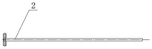
[0028] Example 1 : see Figure 9 , a femoral extramedullary positioning device for knee joint replacement, the positioning device includes an anterior superior iliac spine fixed arch 1 and a first connecting rod 2, the first connecting rod 2 is vertically connected to the anterior superior iliac spine fixed arch 1, The other end of the first connecting rod is connected to the osteotomy positioner 3 , and the positioning device further includes a measuring rod 4 connected to the osteotomy positioner 3 . The overall design of the positioning device is ingenious. The femoral head is positioned with reference to the anterior superior iliac spine on the preoperative plain pelvic radiograph. The extramedullary positioning device is composed of a rigid structure, and the tool can perform multiple measurements, which further improves the positioning accuracy.

Embodiment 2

[0029] Example 2 : see Figure 9 , as an improvement of the present invention, the positioning device further includes a fixing part 5, and the fixing part fixedly connects the first connecting rod 2 and the osteotomy positioner 3. And ensure that the first connecting rod 2 and the osteotomy positioner 3 are perpendicular to each other. The rest of the structures and advantages are exactly the same as in Embodiment 1.

Embodiment 3

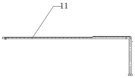
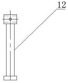
[0030] Example 3 : see figure 1 , 2 , as an improvement of the present invention, the anterior superior iliac spine fixed arch 1 is composed of an L-shaped column and a T-shaped column, and the T-shaped column is sleeved on the crossbar of the L-shaped column, and can be adjusted to the iliac column on both sides. For the length of the anterior superior spine, the position between the center of the femoral head and the vertical foot of the L-shaped crossbar is calculated according to the distance between the anterior superior iliac spine measured by the preoperative pelvic plain film. During the operation, fluoroscopy can also be used to ensure the accuracy of the force line, and the crossbar is marked There are scales to prevent displacement of the connecting rod during the operation. The rest of the structures and advantages are exactly the same as in Embodiment 1.


PUM


Abstract

The invention relates to a femur extramedullary positioning device for knee joint replacement, characterized in that the positioning device includes an anterior superior iliac spine fixation arch (1), a first connecting rod (2), and the first connecting rod (1) It is vertically connected to the fixed arch of the anterior superior iliac spine (1), the other end of the first connecting rod is connected to the osteotomy positioner (3), the positioning device also includes a measuring rod (4), and the measuring rod (4 ) is connected to the osteotomy positioner (3). The advantages of the present invention are as follows: 1) The overall structure is ingeniously designed and easy to install; 2) The technical solution adopts an accurate right-angle connection design and an osteotomy positioning instrument capable of multiple measurements to further ensure its accuracy; 3) The technical solution The handle of the osteotomy locator has complex and fine double fork, groove, scale and small hole design. The double-fork design can constrain the connecting rod in the positioner, and the size is matched to ensure that it is parallel to the connecting rod in the coronal position.

Description

technical field [0001] The invention relates to a positioning device, in particular to a femoral extramedullary positioning device for knee joint replacement, belonging to the field of medical devices. Background technique [0002] Knee replacement is currently widely used clinically. It is a mature surgical technique in joint surgery, and it has an absolute surgical effect on relieving knee joint pain and deformity in elderly patients. However, surgery also brings many risks. Due to the relatively large surgical trauma, post-operative infection, bleeding, and pulmonary embolism have also brought many problems. With the rapid development of knee prostheses today, the key to the efficacy of knee surgery is still the surgical technique, and the accuracy of the force line of knee replacement is closely related to the long-term life of the prosthesis. The tibial positioning system is currently recognized as extramedullary positioning. Because the tibial shaft is relatively supe...

Claims


Application Information

Patent Timeline
Patent Type & Authority Patents(China)
IPC IPC(8): A61F2/46
Inventor 徐志宏蒋青陈东阳
Owner THE AFFILIATED DRUM TOWER HOSPITAL MEDICAL SCHOOL OF NANJING UNIV
Why Eureka
Try Eureka