Double-mode mammary gland three-dimensional imaging device and method

A three-dimensional imaging, dual-mode technology, applied in the field of biomedical imaging, can solve the problems of optical imaging that is difficult to obtain tomographic image reconstruction, difficult to cooperate with, lack of correlation, etc.

Active Publication Date: 2015-03-04
NANJING STARTON MEDICAL TECH CO LTD
View PDF16 Cites 27 Cited by
  • Summary
  • Abstract
  • Description
  • Claims
  • Application Information

AI Technical Summary

Problems solved by technology

However, there are two biggest problems with this technology: one is that the optical image needs external mammography images or ultrasound images to coordinate positioning, so as to find suspicious areas; but because the external image and optical image lack spatial correlation, it is difficult to use them together ; Second, it is difficult to obtain stable tomographic image reconstruction with optical imaging of local fixed point light sources

Method used


Image

  • Double-mode mammary gland three-dimensional imaging device and method
  • Double-mode mammary gland three-dimensional imaging device and method
  • Double-mode mammary gland three-dimensional imaging device and method

Examples


Embodiment approach 1

[0073] Embodiment 1 Three-dimensional imaging device:

[0074] (1) Imaging device:

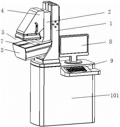
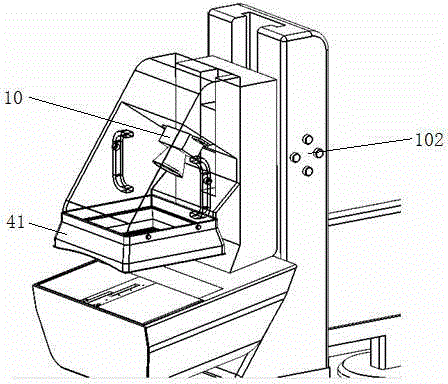
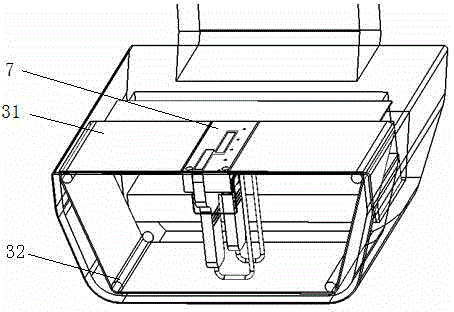
[0075] Such as Figure 1 to Figure 4 As shown, a dual-mode breast three-dimensional imaging device includes a frame 1, a platform 2, a scanning platform 3, a measuring arm 4, a main box 101, a control module 6, a display device 8, a probe scanning mechanism 7, and an opening for placing breasts. 5 and operation keyboard 9. The main frame 101 is arranged on the bottom of the frame 1, the main frame 101 is placed with a main frame, the display device 8 is placed on the table above the main frame 101, and one side of the main frame 101 ( figure 1 An operating keyboard 9 is placed on the protruding horizontal support plate on the front side shown (closer to the side of the user), the display device 8 and the operating keyboard 9 are connected to the host computer, and the host computer and the control module 6 are connected in communication. The frame 1 and the main chassis 101 can also be sepa...

Embodiment approach 2

[0100] Embodiment 2 Three-dimensional imaging method:

[0101] A dual-mode breast three-dimensional imaging method, such as Figure 7 As shown, the three-dimensional imaging device of the present invention is turned on, the display screen lights up, the dual-mode breast three-dimensional imaging software is started, the device performs self-inspection, automatically initializes, the moving parts reset, and the probe returns to the initial position; the user interface is displayed on the display screen, The user sets control parameters through the user interface, such as scanning time or image sampling number; the dual-mode breast three-dimensional imaging method also includes the following steps:

[0102] Adjust the height of the bench 2 to make it suitable for the height of the person to be measured.

[0103] An ultrasonic coupling agent transparent to infrared light is uniformly coated on the upper surface of the scanning sliding film 31 of the scanning platform 2 . The ul...


PUM


Abstract

The invention discloses a novel double-mode mammary gland three-dimensional imaging device and method. The device comprises a main machine body, a rack, a scanning platform, a measuring arm, an ultrasonic imaging unit, an infrared imaging unit, a control module, an infrared imaging device which is mounted in the measuring arm and a probe scanning mechanism which is mounted in the scanning platform, an ultrasonic probe and an infrared light emitting device are disposed on the probe scanning mechanism, and the scanning mechanism is opposite to the infrared imaging device. By means of the double-mode mammary gland three-dimensional imaging device and method, collection of ultrasonic image signals and infrared image signals can be performed simultaneously during one-time scanning, the ultrasonic images are related to the infrared images in space, breast three-dimensional volume ultrasonic images and infrared images can be re-established, overlay analysis of double-mode images can be achieved, check and diagnosis for breast tissue can be facilitated, and noninvasive undamaged and non-radiative effects are achieved.

Description

technical field [0001] The invention belongs to the field of biomedical imaging, and is used for mammography and breast disease detection, in particular to a dual-mode breast three-dimensional imaging device that uses ultrasonic and infrared light synchronously scanning to display three-dimensional breast internal structure ultrasonic images and infrared images, and based on The device's dual-mode 3D imaging approach. Background technique [0002] In mammography (breast) imaging, commonly used detection equipment includes mammography, ultrasound imaging, optical imaging, and magnetic resonance imaging (MRI). Among them, mammography has a wider detection level, simple imaging, and repeatability, and is currently a new standard for breast cancer screening. However, mammography has X-ray radiation, requires a special protective environment, and is not effective for dense breast examination. Ultrasound and magnetic resonance imaging (MRI) also play a complementary role in the ...

Claims


Application Information

Patent Timeline
Patent Type & Authority Applications(China)
IPC IPC(8): A61B5/00A61B8/00
CPCA61B5/0059A61B5/0091A61B5/708A61B8/0825A61B8/40A61B8/4416
Inventor 王维平
Owner NANJING STARTON MEDICAL TECH CO LTD
Why Eureka
Try Eureka