Extraction method of vascular perfusion area in contrast-enhanced ultrasound images based on brox optical flow method

A technology of contrast-enhanced ultrasound and extraction methods, applied in the field of medical image processing, can solve problems that are difficult to apply to clinical data, difficult to coordinate scanning, and complex modeling and analysis processes

Inactive Publication Date: 2017-05-03
THE THIRD AFFILIATED HOSPITAL OF THIRD MILITARY MEDICAL UNIV OF PLA
View PDF2 Cites 0 Cited by
  • Summary
  • Abstract
  • Description
  • Claims
  • Application Information

AI Technical Summary

Problems solved by technology

This imaging method requires the release probe and the imaging probe to focus on the same imaging target area, and it is difficult for the two probes to coordinate scanning, so this method is difficult to achieve successfully; Song Yanchun et al. of Xi'an Jiaotong University also proposed a method based on high mechanical index ( MI), the radio frequency echo decorrelation (DCR) detection method of microbubble destruction caused by single imaging pulse emission, has achieved good experimental results in phantom experiments
[0004] From the above description, it can be found that most of the existing technologies are based on the echo signals generated by destructive imaging to perform decorrelation calculations. The modeling and analysis process is complicated, and it is only implemented in the isolated perfusion phantom model. Difficult to apply to clinical data

Method used


Image

  • Extraction method of vascular perfusion area in contrast-enhanced ultrasound images based on brox optical flow method
  • Extraction method of vascular perfusion area in contrast-enhanced ultrasound images based on brox optical flow method
  • Extraction method of vascular perfusion area in contrast-enhanced ultrasound images based on brox optical flow method

Examples


Embodiment Construction

[0034] The specific implementation manner and working principle of the present invention will be further described in detail below in conjunction with the accompanying drawings.

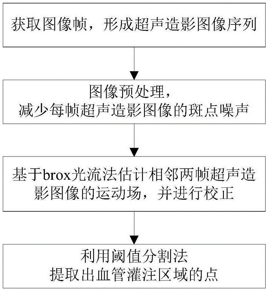
[0035] Such as figure 1 As shown, a method for extracting vascular perfusion area of ​​contrast-enhanced ultrasound images based on brox optical flow method is carried out according to the following steps:

[0036] Step 1: Acquire image frames to form a sequence of contrast-enhanced ultrasound images, specifically:

[0037] Step 1-1: Capture a video of the peak perfusion period of 3 to 8 seconds;

[0038] Step 1-2: Obtain image frames at a sampling rate of 5 frames per second and serially number them;

[0039] Step 1-3: Select images with odd numbers and renumber to form a new image sequence.

[0040] The experimental data in this case comes from patients with uterine fibroids in Chongqing Haifu Hospital. In the implementation process, 2.0ml (10.0mg) of contrast agent Sonovox needs to be injected ...


PUM


Abstract

The invention discloses a method for extracting a blood vessel perfusion region from contrast-enhanced ultrasound images based on a brox optical flow method. The method comprises the steps of (1) obtaining image frames and forming a contrast-enhanced ultrasound image sequence, (2) conducting image preprocessing to reduce speckle noise of each contrast-enhanced ultrasound image, (3) estimating a motion field of every two frames of adjacent contrast-enhanced ultrasound images based on the brox optical flow method, and correcting the two frames of adjacent contrast-enhanced ultrasound images according to an estimated deviation value, and (4) extracting points of the blood vessel perfusion region through a threshold segmentation method. The method has the advantages that the brox optical flow method which is insensitive to noise and high in robustness is adopted to estimate motion displacement of the adjacent image frames and correct the motion displacement; based on the corrected contrast-enhanced ultrasound image sequence, the perfusion region is extracted different from signal features of other tissue backgrounds according to highlight and flashing expressed by radiography microbubbles in blood; experiment results show that the method is small in calculated quantity, easy to realize and good in extraction effect when applied to clinical data.

Description

technical field [0001] The invention relates to a medical image processing technology, in particular to a method for extracting a blood vessel perfusion region of an ultrasound contrast-enhanced image based on brox optical flow method. Background technique [0002] Contrast-enhanced ultrasound imaging is a new type of imaging technology that can dynamically and clearly display tiny blood vessels by injecting contrast agents. This technology can obtain rich blood supply and blood perfusion information of tissues. Contrast microbubbles have strong scattering properties and form a high acoustic impedance difference with the surrounding blood. After being injected into the blood stream, the blood echo can be enhanced to achieve a good "vessel imaging" effect. Extracting the vascular perfusion area from the strong background of the surrounding tissue is the basis of the quantitative analysis of the contrast signal, which has important clinical significance for the clinical diagno...

Claims


Application Information

Patent Timeline
Patent Type & Authority Patents(China)
IPC IPC(8): G06T7/00G06T7/10G06T7/246
CPCG06T7/0012G06T7/10G06T2207/10061G06T2207/10132G06T2207/20104G06T2207/30096G06T2207/30104
Inventor 朱新建吴若愚单鑫吴宝明
Owner THE THIRD AFFILIATED HOSPITAL OF THIRD MILITARY MEDICAL UNIV OF PLA
Why Eureka
Try Eureka