Eureka AIR delivers breakthrough ideas for toughest innovation challenges, trusted by R&D personnel around the world.

Non-linear structure light illumination microscopic imaging method and system

A technology of structured light illumination and microscopic imaging, applied in fluorescence/phosphorescence, material excitation analysis, etc., can solve problems such as limiting NL-SIM, high excitation light power density, biological tissue damage, etc., and achieve improved imaging resolution and imaging. Fast, damage-avoiding effects

Active Publication Date: 2015-04-15
SUZHOU INST OF BIOMEDICAL ENG & TECH CHINESE ACADEMY OF SCI
View PDF4 Cites 44 Cited by
  • Summary
  • Abstract
  • Description
  • Claims
  • Application Information

AI Technical Summary

Problems solved by technology

In 2005, Gustafsson.etal. [PNAS, 102: 13801 (2005)] proposed a nonlinear structured light illumination super-resolution imaging method (NL-SIM), which can obtain high-frequency information of samples by using the saturated nonlinear characteristics of fluorescent molecules , in theory, any high-resolution fluorescence imaging can be achieved. However, in order to achieve saturated nonlinear fluorescence emission, a high excitation light power density is required, which is easy to cause damage to biological tissues. At the same time, NL-SIM needs to collect data that is several times that of SIM. In order to reconstruct a super-resolution image, the imaging speed is slow, these problems limit the practical application of NL-SIM in the field of biomedicine

Method used


Image

  • Non-linear structure light illumination microscopic imaging method and system
  • Non-linear structure light illumination microscopic imaging method and system
  • Non-linear structure light illumination microscopic imaging method and system

Examples


Embodiment Construction

[0035] Preferred embodiments of the present invention will be described in detail below in conjunction with the accompanying drawings.

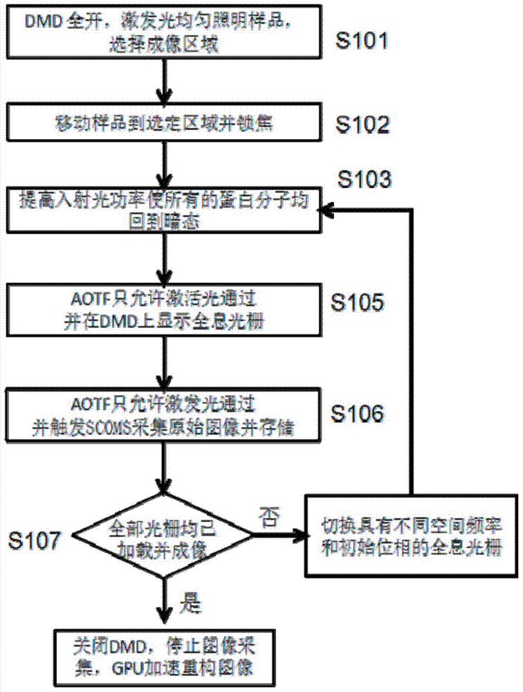
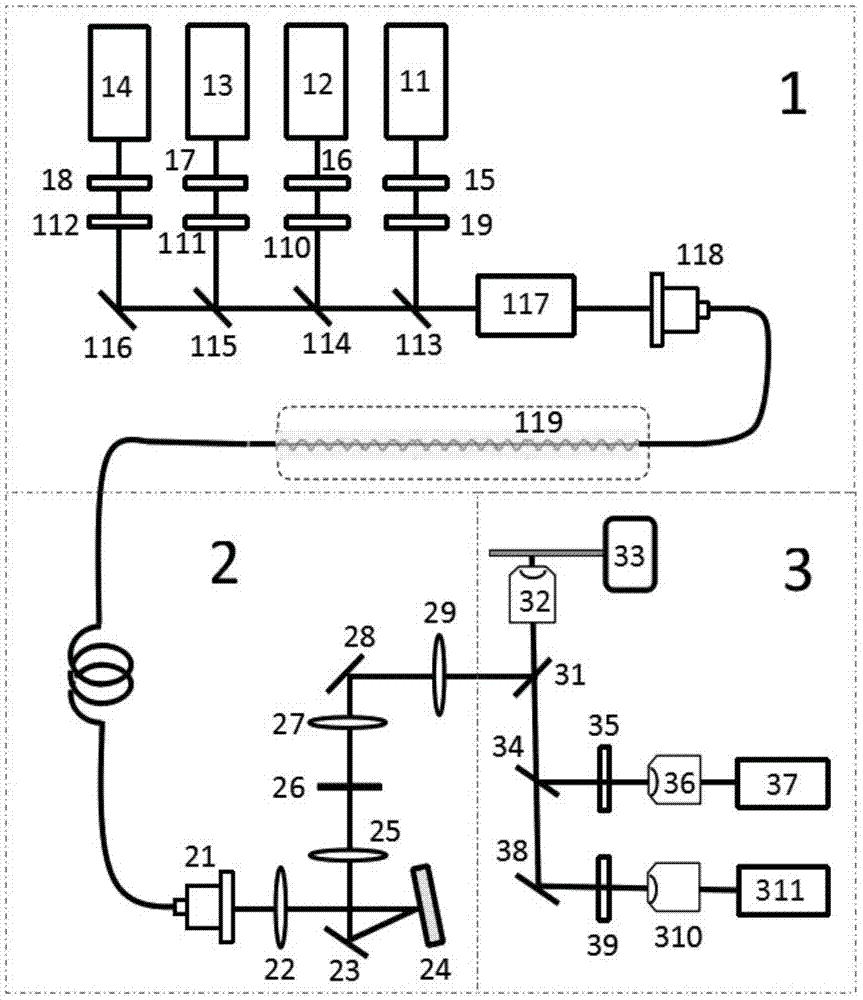
[0036] figure 1 It is a flowchart of a nonlinear structured light illumination super-resolution fluorescence imaging method. For the convenience of description, in this embodiment, the activation light specifically refers to a continuous solid-state laser with a wavelength of 405 nm, and the excitation light specifically refers to a continuous solid-state laser with a wavelength of 488 nm. The output power of the two laser beams is adjustable between 0 and 100 mW.

[0037] In order to achieve the purpose of the present invention, as figure 1 and 3 As shown, in some embodiments of the nonlinear structured illumination microscopy imaging method of the present invention, it includes the steps of:

[0038] Step S101: Uniformly illuminate the sample with excitation light, collect traditional wide-field illumination imaging images through SCOMS,...


PUM


Abstract

The invention discloses a non-linear structure light illumination microscopic imaging method which comprises the following steps: 1) loading a computed hologram on a digital microscopic array; 2) generating a first spatial structure light field which meets sine distribution and is used for activating fluorescent protein, and radiating the first spatial structure light field to the surface of a sample, so as to convert a part of protein to be in an illuminated state from a dark state; 3) radiating the sample in a second spatial structure light field so as to enable fluorescent protein in the bright state to emit fluorescent light, collecting the fluorescent light, and imaging in a photoelectric detector; 4) repeating the step 2) and the step 3), acquiring a plurality of spatial frequencies, acquiring a plurality of initial phases in each direction to obtain a plurality of original images, and reestablishing a super-resolution image according to a GPU acceleration algorithm. The invention further discloses a non-linear structure light illumination microscopic imaging system. The non-linear structure light illumination microscopic imaging method has the advantages of relatively high system imaging resolution, high fluorescent drifting resistance, low phototoxicity and rapid imaging.

Description

technical field [0001] The invention relates to the field of biological imaging, in particular to a non-linear structured illumination microscopic imaging method and system. Background technique [0002] Bioimaging is a frontier field of life science involving multiple disciplines. It is a cutting-edge imaging technology for studying biological functions by "seeing" the interaction of living cells in real time and dynamically at different levels such as molecules, cells, tissues, and living bodies. It has become a breakthrough point for major achievements in life sciences today. [0003] The traditional optical microscope is limited by the optical diffraction limit. The size of the diffraction spot (Airy disk) is about half of the ratio of the wavelength of the light source to the numerical aperture of the microscope objective lens. Generally, the highest imaging resolution is around 200-300nm, and it is impossible to directly observe some How important molecules such as ge...

Claims


Application Information

Patent Timeline
IPC IPC(8): G01N21/64
Inventor 李思黾李辉杨西斌杨光梁永熊大曦
Owner SUZHOU INST OF BIOMEDICAL ENG & TECH CHINESE ACADEMY OF SCI
Why Eureka
Eureka Blog
Learn More