Microscopic analyzing device

A microscopic analysis and atomic force microscopy technology, applied in the field of microscopic analysis, can solve the problem that atomic force microscopy imaging cannot qualitatively analyze samples, and achieve the effect of expanding detection methods

Active Publication Date: 2015-06-03
吉林省恒吉科技有限公司
View PDF3 Cites 9 Cited by
  • Summary
  • Abstract
  • Description
  • Claims
  • Application Information

AI Technical Summary

Problems solved by technology

[0006] The purpose of the present invention is to provide a microanalysis device, which solves the problem that the existing atomic force microscopy imaging cannot qualitatively analyze the sample, and can obtain the molecular composition information of the sampl...

Method used


Image

  • Microscopic analyzing device
  • Microscopic analyzing device
  • Microscopic analyzing device

Examples


Embodiment 1

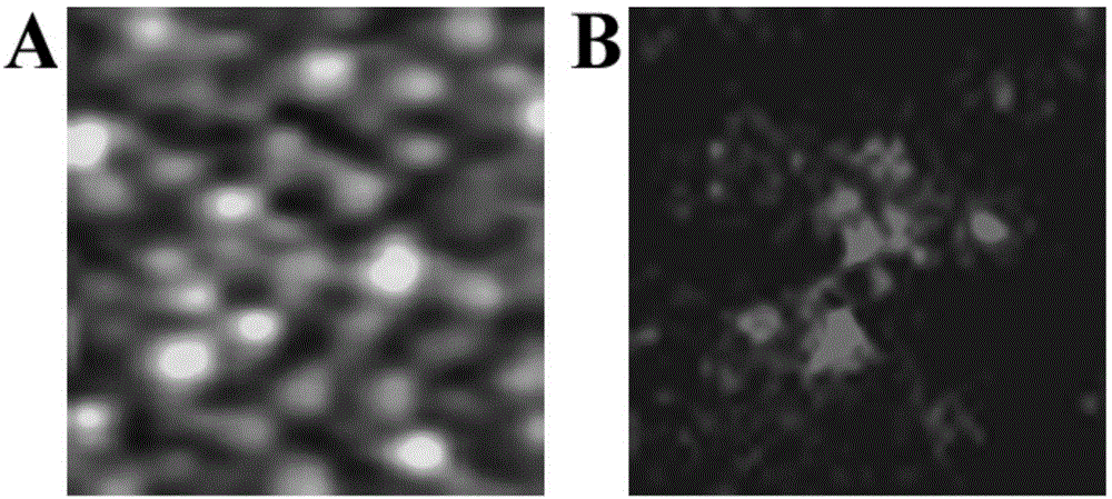
[0052] Conduct high-resolution morphology and composition studies on cell membranes, fix biofilm samples on the sample stage on the surface of the substrate, and perform antibody-labeled staining. The orientation controller in the sample carrying device controls the movement and rotation of the sample stage in three-dimensional directions, so as to select the position of the sample to be measured.

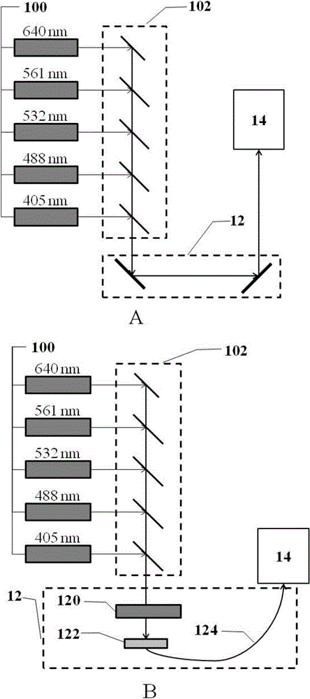
[0053] Five laser emitters respectively emit light with wavelengths of 640nm, 561nm, 532nm, 473nm and 405nm, which are converged into a light source beam by five dichromatic mirrors. The light source beam passes through the optical path attenuator and fiber optic coupler, and is guided by the optical fiber into the random reconstruction fluorescence microscope. The protein in the cell membrane sample is optically positioned by the random reconstruction fluorescence microscope, and the imaging result is output through the computer. Simultaneously with fluorescence imaging, atomic fo...


PUM


Abstract

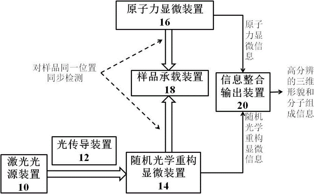
The invention provides a microscopic analyzing device. The microscopic analyzing device comprises a laser source device, a light transmission device, a stochastic optical reconstruction microscopic device, an atomic force microscopic device, a sample bearing device and an information integrating and outputting device. A light source beam provided by the laser source device is guided into the stochastic optical reconstruction microscopic device through the light transmission device, so that a sample to be detected on the sample bearing device is subjected to optical reconstruction microscopic imaging; simultaneously, the atomic force microscopic device carries out atomic force microscopic imaging at the same position; the information integrating and outputting device integrates and outputs stochastic optical reconstruction microscopic information and atomic force microscopic information; therefore, the problem that the sample cannot be qualitatively analyzed by the existing atomic force microscopic imaging can be solved; and thus, the microscopic analyzing device can be widely applied in the fields of biology, polymers and materials.

Description

technical field [0001] The invention relates to a combination technology of a stochastic optical reconstruction micro-device and an atomic force micro-device, belonging to the field of microanalysis. Background technique [0002] Optical microscope is an essential tool for biologists to do research and explore the mysteries of life. Light microscopy has been used to gain a first glimpse at the intricate structure and morphology inside cells. However, limited by the optical diffraction limit, the resolution of conventional optical microscopes is difficult to reach below 200nm. In recent years, the invention of super-resolution fluorescence microscopy breaks through the optical diffraction limit. These include stimulated emission depletion microscopy (STED), saturated structured illumination microscopy (SSIM), stochastic optical reconstruction microscopy (STORM), and photoactivated localization microscopy (PALM). The three-dimensional spatial resolution of these methods is ...

Claims


Application Information

Patent Timeline
IPC IPC(8): G01N21/64G02B21/36
Inventor 吴宗斌
Owner 吉林省恒吉科技有限公司
Why Eureka
Try Eureka