
Manufacturing method for individual three-dimensional auricle guide plate
A technology of a guide plate and auricle, which is applied in the field of medical image processing, can solve the problems of lengthy, cumbersome operation steps, unfavorable for general doctors to grasp and popularize, etc., and achieves the effect of improving validity and increasing accuracy
- Summary
- Abstract
- Description
- Claims
- Application Information
AI Technical Summary
Problems solved by technology
Method used
Image

Examples
Embodiment 1
[0025] Example 1: Preparation of personalized three-dimensional auricle guide plate for patients with microtia
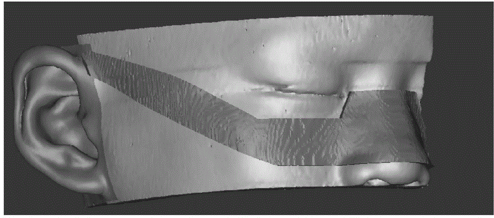
[0026] Firstly, import the DICOM format data obtained from CT scanning into Mimics software, use the midsagittal plane and guide plate positioning, and combine Mimics software to segment, extract, and reconstruct the image data to directly and quickly generate a 3D digital model of the auricle and guide plate on the affected side. Connect it with a 3D printer to create a three-dimensional solid model. Specifically include steps:
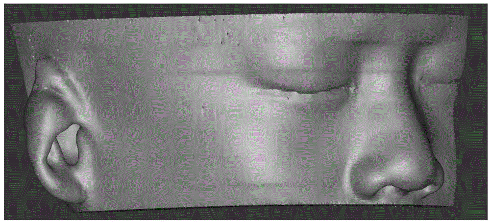
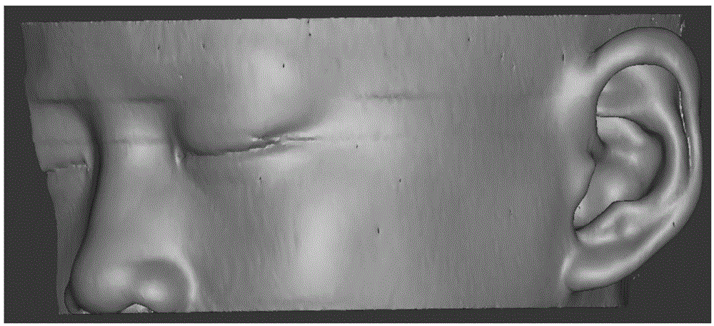
[0027] according to figure 1 , 2 As shown, import the DICOM format data obtained from the temporal bone CT scan of microtia patients into Mimics software, create a new Mask, select the threshold value range from -435 to 330, define the soft tissue, and extract the three-dimensional information of the auricle and face on the healthy side; make a cockscomb The mid-sagittal plane of the apex, the midpoint of the posterior edge of the foram...
PUM
 Abstract
Description
Claims
Application Information