

Pelvic fracture microinvasive intramedullary fixation device
A fixation device and intramedullary nail technology, applied in the field of orthopedic medical devices, to achieve the effects of flexible manipulation, promotion of fracture healing, and less periosteal peeling
- Summary
- Abstract
- Description
- Claims
- Application Information
AI Technical Summary
Problems solved by technology
Method used
Image

Examples
Embodiment Construction
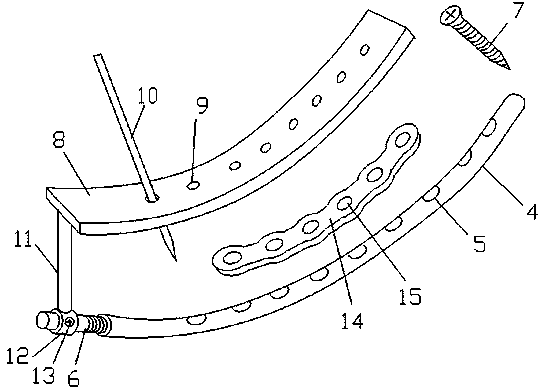
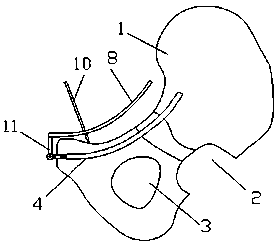
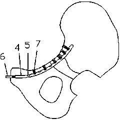
[0020] The present invention includes a guide wire, a guide sleeve, a soft drill, an arc-shaped intramedullary nail 4, an arc-shaped screw guide plate 8, a guide pin 10, a fixing screw 7, an intramedullary nail tail cap 6, a connecting rod 11, and a reconstruction plate 14.
[0021] The guide wire and the guide sleeve of the present invention are curved arcs, the arc matches the radian of the preset pelvic intramedullary tunnel, the outer diameter of the guide sleeve matches the preset inner diameter of the pelvic intramedullary tunnel, and is soft. The drill is located in the guide sleeve, and the guide wire is threaded on the central axis of the soft drill. During the operation, firstly, the soft drill drills the pelvic intramedullary tunnel on the pelvis according to the preset pelvic intramedullary tunnel path under the guidance of the guide wire and the guide sleeve, and then puts the arc-shaped intramedullary nail 4 in the tunnel, Immobilize the fracture site.
[0022] ...
PUM
 Abstract
Description
Claims
Application Information