Automatic fast segmenting method of tumor pathological image

A technology for pathological images and tumors, which is applied in the field of image processing and can solve the problems of high algorithm complexity, difficulty in accurate segmentation, and long processing time.

Active Publication Date: 2015-09-23
NANTONG UNIVERSITY
View PDF2 Cites 34 Cited by
  • Summary
  • Abstract
  • Description
  • Claims
  • Application Information

AI Technical Summary

Problems solved by technology

These methods have some major disadvantages: (1) Most methods require manual interaction; (2) Most methods are sensitive to noise, and due to the low contrast of ultrasound images and tissue-related textures, accurate segmentation is difficult; (3) Most methods The complexity of the algorithm is high, the processing time is long, and it is difficult to meet the clinical requirements
However, these methods have the following problems: (1) The value of multi-resolution

Method used


Image

  • Automatic fast segmenting method of tumor pathological image
  • Automatic fast segmenting method of tumor pathological image
  • Automatic fast segmenting method of tumor pathological image

Examples


Embodiment 1

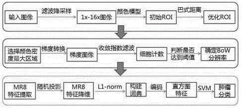
[0058] An accurate, fast and automatic segmentation method for tumor pathological images. In order to combine clinician's experience and knowledge with image processing technology, firstly, Gaussian pyramid algorithm is used to filter the original tumor pathological images, and the results are respectively obtained from 1 times, 2x, 4x, 8x, 16x pathological images, through the RGB color model and morphological "close operation" to determine the initial region of interest containing the tumor on the 1x resolution image; The iterative optimization of the initial tumor ROI was carried out from 1X resolution to 4X resolution. When the Basset distance reached a certain threshold, it was judged that the contribution of the RGB color model to the tumor ROI had been reduced to zero. Then use the convergence index filter algorithm for adaptive high-resolution selection of depth-accurate segmentation, so as to perform further segmentation at the most suitable high-resolution; finally, us...

Embodiment 2

[0091] The method in Embodiment 1 is compared with the VZ_MR8 and TFISF methods in terms of encoding time. (Both VZ_MR8 and TFISF methods are obtained by referring to the paper "Effective texture classification by texton encoding induced statistical features") KTH_TIPS is an open source database of texture images (available at www.nada.kth.se / cvap / databases / kth-tips / ) , including 10 types of images, each type of image includes 81 pictures, and each image size is 200×200 pixels. The Medical block database is based on 120 colorectal tumor pathological slice images according to the manual identification of clinicians, randomly extracting 1000 8-fold and 16-fold patches each, and each patch is 200×200 pixels in size, including 500 8-fold tumor patches, 8 times normal tissue patches500, 16 times tumor patches500 pieces, 16 times normal tissue patches500 pieces. The experimental results are the average of 20 experiments, as shown in Table 1. The encoding time based on the random p...


PUM


Abstract

The invention discloses an automatic fast segmenting method of a tumor pathological image. The method comprises the following steps: firstly filtering a tumor original pathological image through the adoption of a Gaussian pyramid algorithm to respectively obtain pathological images with equal resolution, double resolution, fourfold resolution, eightfold resolution and 16-fold resolution; determining an initial region of interest containing the tumor on the equal resolution image through a RGB color model and morphological close operation; iteratively optimizing the initial regions of interest from the equal resolution to the fourfold resolution through the adoption of bhattacharyya distance; judging that the contribution of the RGB color model to the tumor region of interest has been reduced to zero when the bhattacharyya distance achieves a set threshold value; performing the self-adaptive high resolution selection of the deep precise segmentation through the adoption of a convergence exponent filtering algorithm, thereby further segmenting under the most suitable high resolution; and finally segmenting out a normal tissue and a tumor tissue in the tumor region of interest through the adoption of a bag of words model based on random projection. The method disclosed by the invention has the features of being accurate, fast and automatic.

Description

technical field [0001] The invention belongs to the technical field of image processing, and in particular relates to a method for automatic and fast segmentation of tumor pathological images. Background technique [0002] Cancer has become one of the leading causes of death in developed countries. Taking colorectal cancer as an example, it is currently the third largest malignant tumor in the world. Due to the characteristics of full color and high image resolution, the current pathological section diagnosis has become one of the important means of tumor detection. However, at present, tumor diagnosis is completely dependent on the manual operation of pathologists, which is not only slow in efficiency, but also highly affected by the subjective judgment of doctors. Therefore, the computer-aided diagnosis system based on pathology is of great significance. Some studies have shown that the output results of pathology computer-aided diagnosis system can provide a reference fo...

Claims


Application Information

Patent Timeline
IPC IPC(8): G06T7/00
CPCG06T7/11G06T2207/20081G06T2207/20104G06T2207/30096
Inventor 张堃吴建国张培建杨晓伟顾磊楚启超
Owner NANTONG UNIVERSITY
Why Eureka
Try Eureka