Cerebrovascular image segmentation method based on multi-angle serialized image space feature point set

A feature point set and image space technology, applied in the field of image processing, can solve problems affecting doctors' diagnosis, large noise in blood vessel images, and time-consuming problems

Active Publication Date: 2015-10-28
DALIAN UNIV OF TECH
View PDF7 Cites 17 Cited by
  • Summary
  • Abstract
  • Description
  • Claims
  • Application Information

AI Technical Summary

Problems solved by technology

The manual segmentation method requires manual interaction, the segmentation results obtained by different manual operations are very different, and it takes a lot of time, and the blood vessel images obtained by the registration method contain large noise, which affects the doctor's diagnosis. The model method requires a template image, and only an accurate template can get better blood vessel segmentation results

Method used


Image

  • Cerebrovascular image segmentation method based on multi-angle serialized image space feature point set
  • Cerebrovascular image segmentation method based on multi-angle serialized image space feature point set
  • Cerebrovascular image segmentation method based on multi-angle serialized image space feature point set

Examples


Embodiment Construction

[0080] In order to make the technical solutions and advantages of the present invention more clear, the technical solutions in the embodiments of the present invention are clearly and completely described below in conjunction with the drawings in the embodiments of the present invention:

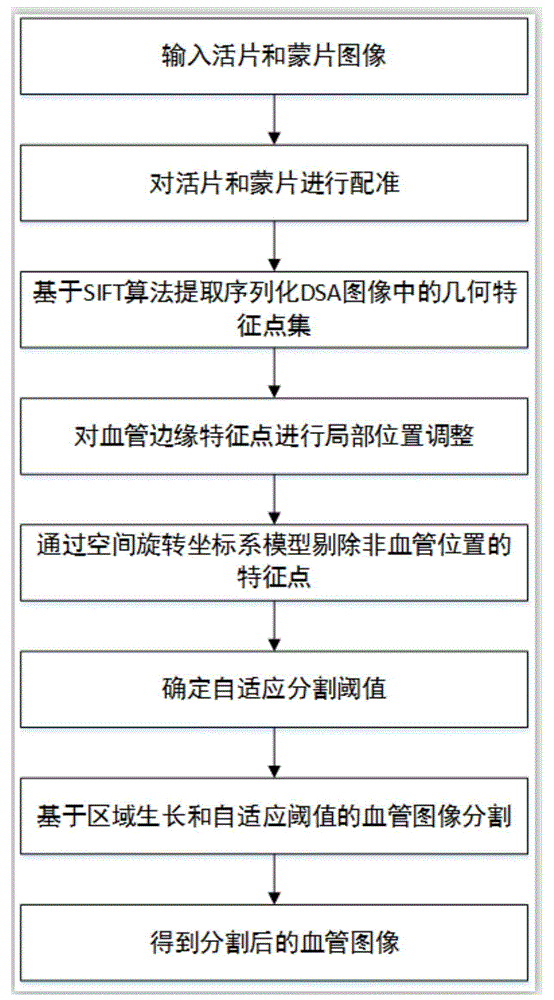
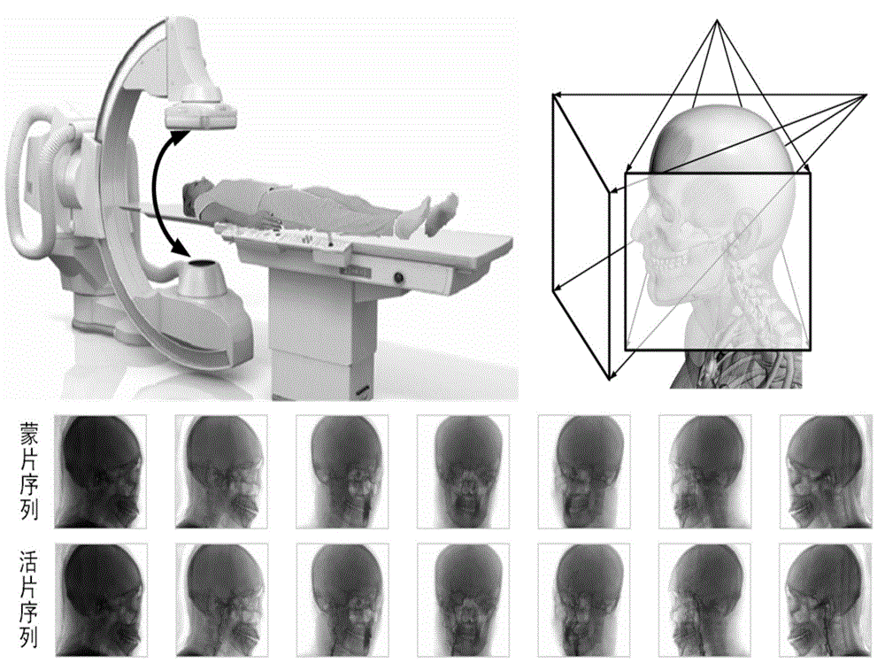
[0081] Such as figure 1 A cerebrovascular image segmentation method based on a multi-angle serialized image space feature point set is shown. In the implementation process, the schematic diagram of the image sequence acquisition process is as follows figure 2 , the upper left corner is the image acquisition device, and the upper right corner is a schematic diagram of collecting DSA image sequences from different angles. The equipment rotates the C-arm to obtain mask and live image sequences after acquisition, and then performs "pair by pair" processing to extract feature point sets. Then introduce the rotating coordinate system to remove dirty data, and finally perform segmentation processi...


PUM


Abstract

The invention discloses a cerebrovascular image segmentation method based on a multi-angle serialized image space feature point set, comprising the following steps: S1, registering a live image and a mask image of cerebral vessels; S2, extracting a geometric feature point set of a serialized subtraction image; S3, locally adjusting the positions of the feature points at the edges of the blood vessels in the serialized subtraction image; S4, adopting a spatial rotating coordinate system to remove the feature points in non-vascular positions in the subtraction image; S5, determining the adaptive segmentation threshold of the serialized subtraction image; and S6, based on region growing and vascular image segmentation of adaptive threshold and by taking the feature points obtained in S4 as seed points and the adaptive segmentation threshold obtained in S5 as the standard of the law of growth, adopting a region growing algorithm to segment the serialized subtraction image to obtain a pure cerebrovascular image.

Description

technical field [0001] The invention relates to the technical field of image processing, in particular to a method for segmenting cerebrovascular images based on multi-angle serialized image space feature point sets. Background technique [0002] Obtaining images of cerebral blood vessels through DSA technology can help doctors diagnose patients, but due to the patient's breathing, swallowing and other actions, there will be obvious artifacts and noises, which will affect the doctor's observation and diagnosis. Therefore, accurate vascular tissue extraction Image is an urgent problem to be solved in the current vascular diagnosis and treatment based on DSA technology. Existing techniques include manual segmentation methods, registration methods, and model-based methods. The manual segmentation method requires manual interaction. The segmentation results obtained by different manual operations are very different, and it takes a lot of time. The blood vessel images obtained b...

Claims


Application Information

Patent Timeline
Patent Type & Authority Applications(China)
IPC IPC(8): G06T7/00
CPCG06T2207/10
Inventor 刘斌江乾峰黄睿刘文鹏
Owner DALIAN UNIV OF TECH
Why Eureka
Try Eureka