
Separation method for megakaryocyte progenitors

A separation method and technology of nuclear cells, applied in animal cells, vertebrate cells, blood/immune system cells, etc., can solve the problems of low megakaryocyte ratio and low yield

Inactive Publication Date: 2016-01-20
GUANGZHOU SALIAI STEMCELL SCI & TECH CO LTD
View PDF3 Cites 2 Cited by
  • Summary
  • Abstract
  • Description
  • Claims
  • Application Information

AI Technical Summary

Problems solved by technology

The method provided by the former is relatively simple, but the yield is low; the yield of the latter is relatively high, but the method provided focuses on obtaining megakaryotic progenitor cells, but ignores its prerequisites. The number of isolated mononuclear spheres directly affects the megakaryotic progenitor cells. The yield, and the methylcellulose used in the separation process of the method cannot be used for human intravenous infusion, and the resulting megakaryocyte progenitor cells induce a low ratio of megakaryocytes (CD41a+ / CD61+ cells)

Method used


Image

  • Separation method for megakaryocyte progenitors

Examples


Embodiment 1

[0038] Example 1 Mononuclear cell isolation

[0039] Add 25 mL of hydroxyethyl starch injection (Beijing Shuanghe Pharmaceutical Co., Ltd.) (cord blood: hydroxyethyl starch = 2:1 (v / v)) to 50 mL of the collected cord blood, mix well, and stand still for 15 minutes Above, sediment erythrocytes, and aspirate the supernatant;

[0040] The supernatant was added to a centrifuge tube containing lymphocyte separation solution (supernatant:separation solution=2:1 (v / v)), centrifuged at 3000 rpm for 20 min, and mononuclear cells were collected.

[0041] The collected mononuclear cells were washed with PBS and centrifuged at 1500 rpm for 10 min, repeated twice. After cell counting, the number of mononuclear cells was 5.82×10 7 , of which the number of CD34+ cells is 3.05×10 5 indivual.

Embodiment 2

[0042] Example 2 Mononuclear cell isolation

[0043] Add 12.5 mL of hydroxyethyl starch injection (Beijing Shuanghe Pharmaceutical Co., Ltd.) (cord blood: hydroxyethyl starch = 4:1 (v / v)) to 50 mL of collected umbilical cord blood, mix well, stand still More than 15min, sediment erythrocytes, suck the supernatant;

[0044] The supernatant was added to a centrifuge tube containing lymphocyte separation solution (supernatant:separation solution=2:1 (v / v)), centrifuged at 3000 rpm for 20 min, and mononuclear cells were collected.

[0045] The collected mononuclear cells were washed with PBS and centrifuged at 1500 rpm for 10 min, repeated twice. After cell counting, the number of mononuclear cells was 6.15×10 7 of which the number of CD34+ cells is 3.18×10 5 indivual.

Embodiment 3

[0046] Example 3 Mononuclear cell isolation

[0047] Add 8.3 mL of hydroxyethyl starch injection (Beijing Shuanghe Pharmaceutical Co., Ltd.) (cord blood: hydroxyethyl starch = 6:1 (v / v)) to 50 mL of the collected cord blood, mix well, and stand still. More than 15min, sediment erythrocytes, suck the supernatant;

[0048] The supernatant was added to a centrifuge tube containing lymphocyte separation solution (supernatant:separation solution=2:1 (v / v)), centrifuged at 3000 rpm for 20 min, and mononuclear cells were collected.

[0049] The collected mononuclear cells were washed with PBS and centrifuged at 1500 rpm for 10 min, repeated twice. After cell counting, the number of mononuclear cells was 6.04×10 7 of which the number of CD34+ cells is 3.10×10 5 indivual.


PUM


Abstract

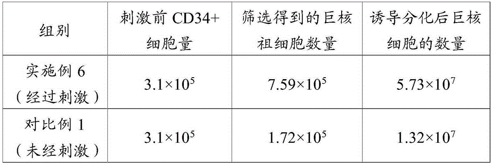
The invention relates to the field of cell culture, in particular to a separation method for megakaryocyte progenitors. According to the method, stimulation culturing is performed on single karyocytes obtained through separation through rhG-CSFs, so that the content of megakaryocytes in the cells is increased, the cells processed through stimulation culturing are screened with immunomagnetic beads, and the megakaryocyte progenitors are obtained. The megakaryocyte progenitors are isolated by adopting the method, the yield is higher, and the number of the megakaryocytes (the phenotype is CD41a<+> / CD61<+>) obtained through induced directional differentiation is larger. Experiments show that 20-100 mL of umbilical cord blood is separated through the method, the number of the obtained megakaryocyte progenitors can reach 2.75*10<6>, the quantity percentage of the megakaryocytes in the obtained cells is 5%-10%, and after 14 days of induced directional differentiation, the total number of the megakaryocytes can be amplified by 132.5+ / -21.2 times, and the proportion of the megakaryocytes can reach 81%.

Description

technical field [0001] The invention relates to the field of cell culture, in particular to a method for separating megakaryotic progenitor cells. Background technique [0002] Megakaryocyte progenitor cells (megakaryocyte progenitor cells) are a kind of cells that make people produce megakaryocytes, which are derived from bone marrow progenitors (CFU-GEMM), referred to as megakaryocyte progenitor cells. Megakaryotic progenitor cells are present on CD126 - / Lin - Among the cell populations, CD34+ cells can be divided into two cell subsets, expressing and not expressing IL-6R, IL-6R+ cells can be stimulated to form granulocytes-macrophages, lymphocytes and granulocytes, while IL-6R+ cells can be stimulated to form granulocytes, lymphocytes and granulocytes. 6R-cells can form erythroid and megakaryotic cells when administered IL-6 / sIL-6R. Hematopoietic stem cells first differentiate into megakaryoid progenitor cells, also known as colony forming unit-megakaryocytes (CFU-Meg...

Claims


Application Information

Patent Timeline
IPC IPC(8): C12N5/0789
Inventor 陈海佳王一飞葛啸虎李丽娟万桦
Owner GUANGZHOU SALIAI STEMCELL SCI & TECH CO LTD
Learn More