
X-ray protection device applied to angiography machine

A protective device and imaging machine technology, which is applied in the field of medical equipment, can solve problems such as inconvenient operation, limited protection, and poor practicability, and achieve the effects of reducing external expansion, good barrier effect, and operational effect

Inactive Publication Date: 2016-01-27
王铮
View PDF12 Cites 3 Cited by
  • Summary
  • Abstract
  • Description
  • Claims
  • Application Information

AI Technical Summary

Problems solved by technology

At present, when clinically performing cardiac interventional operations, doctors perform intubation, angiography, filming, drug injection, and stent placement under the guidance of X-rays. A radiation protection device is set up, and the protection device is generally made of materials with a lead equivalent of 0.5mm or more to block X-ray radiation. In the irradiated area, it brings great X-ray damage to the surgeon
Although the device has multiple degrees of freedom to rotate to protect the medical staff, it still cannot use the baffle to protect the position close to the operating bed. Although it is easy to adjust, it has limitations in protection
[0004] The Chinese invention patent titled an integrated X-ray protection device on an angiography machine tool (publication number CN103385733B) discloses an integrated X-ray protection device on an angiography machine tool, which is characterized in that the device includes A protective screen unit for radiation protection above the catheter bed, a protective curtain unit for radiation protection below the catheter bed, and a bedside slide rail unit for connecting side rails beside the catheter bed, wherein: the protective screen unit is installed on On the protective curtain unit, the protective curtain unit is fixed on the bedside slide rail unit, so that the protective curtain unit and the protective screen unit can move horizontally along the bedside synchronously; the protective screen unit includes A rotating arm, a planar hinge four-bar mechanism, a transparent protective screen installed above the rotating arm, and a lifting protective curtain suspended below the rotating arm, wherein: the protective screen unit is hinged on the rotating arm and the planar hinge four-bar mechanism on the protective curtain unit, so that the protective screen unit can rotate horizontally with the rotating arm, and, when the operation state and the protective screen unit is parallel to the protective curtain unit, the protective screen unit is raised to Above the protective curtain unit, when the operation is over, the protective screen unit is lowered and restored to be parallel to the protective curtain unit through a planar hinge four-bar mechanism; the integrated X-ray protection device structure in this scheme is still relatively complicated, and the barrier is not too large. Well, the operation is inconvenient and the practicability is relatively poor

Method used


Image

  • X-ray protection device applied to angiography machine
  • X-ray protection device applied to angiography machine
  • X-ray protection device applied to angiography machine

Examples


Embodiment approach

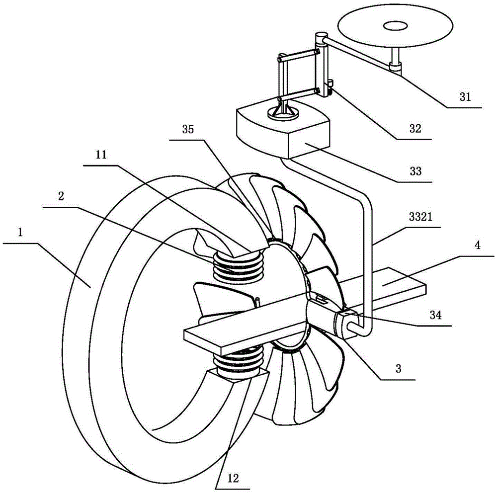
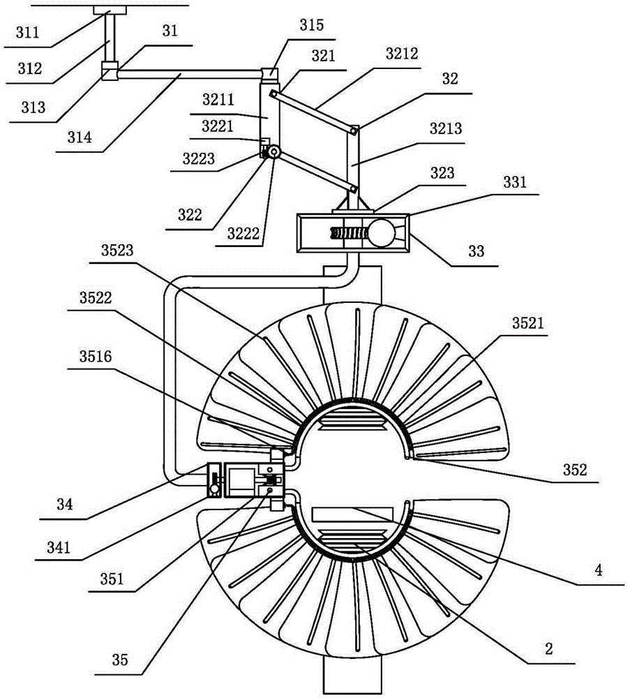
[0034] The present invention provides an embodiment of the fixed rotation unit 33, the fixed rotation unit 33 includes a first support 311, a first steel pipe 312, a first rotating shaft 313, a second steel pipe 314 and a second rotating shaft 315, the upper end of the first steel pipe 312 The first support 311 is fixedly connected to the wall, the lower end of the first steel pipe 312 is connected to one end of the second steel pipe 314 through the first rotating shaft 313, and the other end of the second steel pipe 314 is connected to the lifting unit 32 through the second rotating shaft 315 Above: the fixed rotation unit 33 fixes the shutter mechanism 3, and the fixed rotation unit 33 enables it to be adjusted and rotated to better block X-rays.

[0035] The present invention provides an embodiment of the lifting unit 32. The lifting unit 32 includes a hinged four-bar connection mechanism 321 and a first worm gear mechanism 322; the lifting unit 32 is connected to the first ...


PUM


Abstract

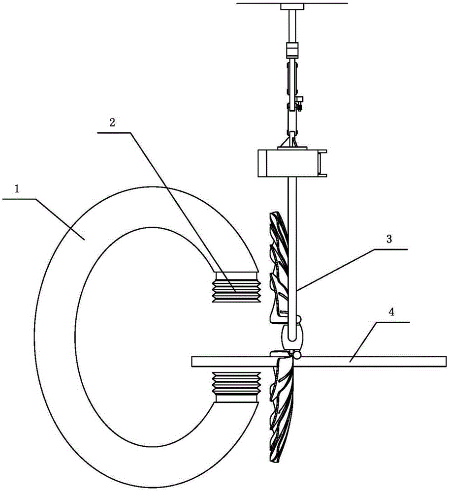
The invention relates to the field of medical apparatuses and instruments, in particular to an X-ray protection device applied to an angiography machine. The X-ray protection device applied to the angiography machine comprises a telescopic pipe and a blocking piece mechanism. In the X-ray protection device applied to the angiography machine, the X-ray machine emits X-rays from an emitting port, the X-rays pass through an operating table and are received by a receiving port, and when the X-rays are emitted from the emitting port, very big outward diffusion phenomena are produced due to the distance between the emitting port and receiving port. Therefore, the emitting port and the receiving port are respectively connected with the telescopic pipe, the telescopic pipe guides a route of the X-rays, the outward X-ray diffusion phenomena are greatly decreased, and accordingly injury to a medical worker is avoided. The blocking piece mechanism is located in the front of the X-ray machine, can be move relative to the X-ray machine and block X-ray refraction on a patient and outward X-ray radiation, and accordingly the injury to the medical worker is avoided. The X-ray protection device is simple in structure, convenient to operate, good in blocking effect and good in practicability.

Description

technical field [0001] The invention relates to the field of medical equipment, in particular to an X-ray protection device applied to an angiography machine. Background technique [0002] As we all know, with the continuous development of modern medicine, cardiac interventional surgery has become one of the important means of treating heart diseases. Catheter intervention, coronary angiography, and stent placement under X-rays can be performed accurately and quickly, and become an important method for timely diagnosis and treatment of patients with myocardial infarction and dissecting aneurysm and to improve the cure rate. In some clinical operations such as angiography and interventional therapy, it is necessary to rely on x-rays penetrating the patient's body to form an image through an angiography machine; in order to avoid leakage of x-ray radiation and protect the safety of medical personnel, radiation protection must be installed next to the angiography machine devic...

Claims


Application Information

Patent Timeline
Patent Type & Authority Applications(China)
IPC IPC(8): A61B6/10
Inventor 王铮孙奇勋王富昌杨洁琼
Owner 王铮
Learn More