Scanning dark field laser speckle blood flow imaging method and device

A technology of laser speckle and imaging method, which is applied in medical science, diagnosis, diagnostic recording/measurement, etc., can solve the problem of low degree of improvement, and achieve the effect of improving detection depth and increasing contribution

Active Publication Date: 2016-03-30
HUAZHONG UNIV OF SCI & TECH
View PDF4 Cites 13 Cited by
  • Summary
  • Abstract
  • Description
  • Claims
  • Application Information

AI Technical Summary

Problems solved by technology

The combination of laser speckle blood flow imaging and orthogonal polarization method can eliminate the specular reflection light on the tissue surface during imaging, thus improving the sampling depth of blood flow signals to a certain extent, but the degree of improvement is not high

Method used


Image

  • Scanning dark field laser speckle blood flow imaging method and device
  • Scanning dark field laser speckle blood flow imaging method and device
  • Scanning dark field laser speckle blood flow imaging method and device

Examples


Embodiment Construction

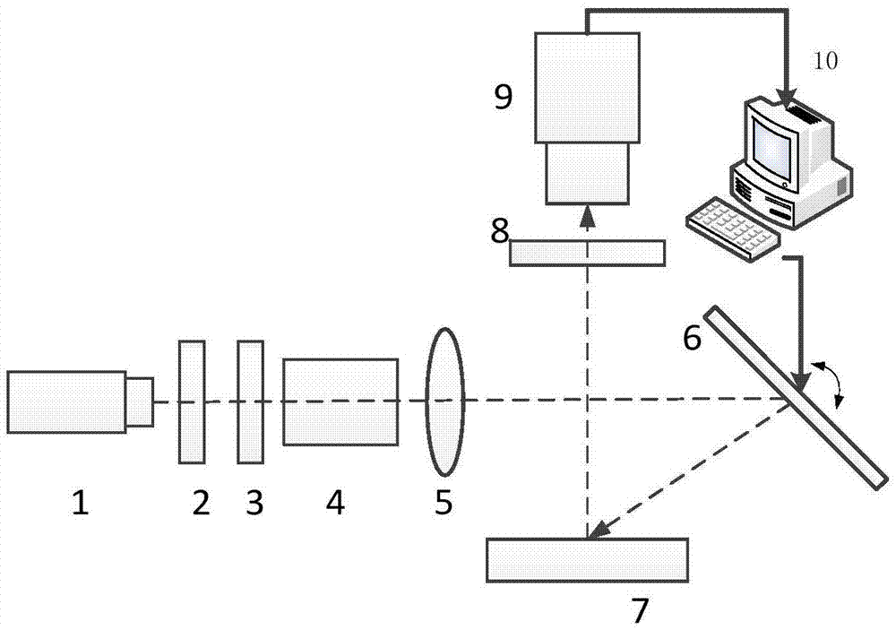
[0029] like figure 1 As shown, the device for scanning dark-field laser speckle blood flow imaging method includes: a laser light source 1, a polarizer 2, a beam shaper 3, a beam expander 4, a cylindrical lens 5, a scanning galvanometer 6, and a sample 7 are located in sequence The illumination optical path, and the polarizer 2 is perpendicular to the incident laser beam 1; the sample 7, the analyzer 8, and the photoelectric imaging system 9 are located on the imaging optical path in turn, and the analyzer 8 is perpendicular to the optical axis of the photoelectric imaging system 9, and the photoelectric imaging system 9 is perpendicular to the photoelectric imaging system. The imaging system 9 is concentric, and its polarization direction is perpendicular to the polarization direction of the analyzer 7; the computer 10 is connected to the scanning galvanometer 6, and the scanning galvanometer is controlled to irradiate the localized laser beam onto the measured object, and sca...


PUM


Abstract

The invention discloses a scanning dark field laser speckle blood flow imaging method and device. Biological tissue is illuminated by means of space localized linear or dotted laser beams, scanning is conducted along the surface of the tissue to be tested, and the whole area to be tested is traversed. During each time of scanning, a laser speckle image reflected by the whole area to be tested (containing the area directly illuminated by the laser beams and the surrounding area not directly illuminated) is acquired by an area array CCD or CMOS camera through an optical imaging system, the laser speckle contrast corresponding to each pixel is calculated, and then the laser speckle image is converted into a blood flow image. Pixel weighted averaging is conducted on the blood flow images obtained by scanning the illumination laser beams at different positions, so that the final two-dimensional blood flow distribution image of the biological tissue in the area to be tested is obtained. The method and device have the advantages that the contribution of diffusion photons subjected to multiple scattering in the tissue to imaging is expanded, and detection depth is improved compared with a traditional laser speckle blood flow imaging method adopting broad-beam laser illumination.

Description

technical field [0001] The invention relates to a scanning dark field laser speckle blood flow imaging method and device, which is used to improve the detection depth of the laser speckle blood flow imaging technology, and is used for physiology, pathology, pharmacology and drug effect evaluation research, as well as clinical medicine Diagnosis and treatment. Background technique [0002] The existing laser speckle blood flow imaging is a wide-field blood flow imaging technology with high temporal and spatial resolution, and has been widely used in basic life science research and clinical disease diagnosis and treatment. However, the sampling depth of this technique is limited, and it mainly detects the blood flow information in the superficial layer of biological tissue. It has been reported that methods to improve the sampling depth of laser speckle blood flow imaging include the use of optical transparent agents, transmission imaging, and the combination of laser speckle...

Claims


Application Information

Patent Timeline
Patent Type & Authority Applications(China)
IPC IPC(8): A61B5/00
Inventor 李鹏程骆清铭何珩唐颖
Owner HUAZHONG UNIV OF SCI & TECH
Why Eureka
Try Eureka