Automatic detection method for microaneurysm in color eye fundus image

A fundus image and automatic detection technology, applied in image enhancement, image analysis, image data processing, etc., can solve problems such as low contrast, uneven illumination, interference, etc., achieve high accuracy, reduce false detection, and strong applicability Effect

Inactive Publication Date: 2016-04-13
TIANJIN POLYTECHNIC UNIV
View PDF0 Cites 9 Cited by
  • Summary
  • Abstract
  • Description
  • Claims
  • Application Information

AI Technical Summary

Problems solved by technology

[0003]However, in the imaging of color fundus images, due to the influence of objective factors such as shooting conditions, human eye structure and different degrees of fundus lesions, uneven illumination generally exists in fundus images , low contrast and much noise
At the same time, due to the small size of the microaneurysm and the low contrast with the local tissue background, the difficulty of microaneurysm detection is greatly increased.
At present, the detection methods for microaneurysms have achieved good results, but the detection results are easily disturbed by small blood vessels and background noise, which reduces the accuracy of the detection method

Method used


Image

  • Automatic detection method for microaneurysm in color eye fundus image
  • Automatic detection method for microaneurysm in color eye fundus image
  • Automatic detection method for microaneurysm in color eye fundus image

Examples


Embodiment Construction

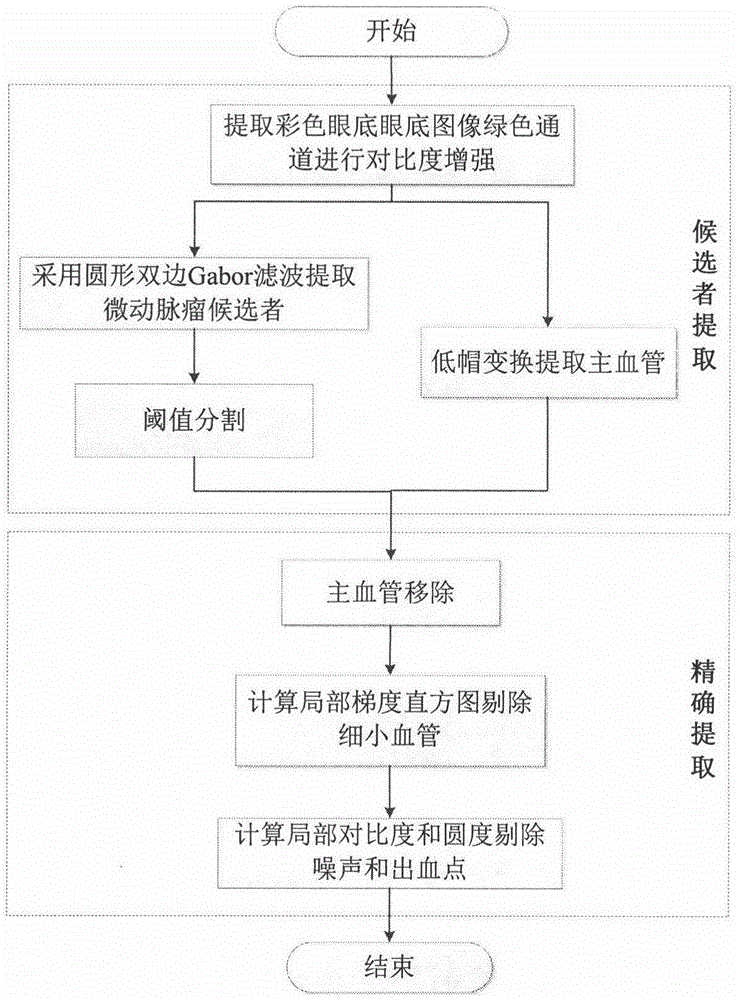
[0026] Process of the present invention Such as figure 1 As shown, the method first uses the contrast enhancement algorithm to analyze the color fundus picture The green channel of the image is preprocessed to enhance the contrast; then low-hat transformation is used to extract the main blood vessels of the fundus, and the circular bilateral Gabor filter and threshold segmentation are used to extract microaneurysm candidates; then the local gray level centered on the candidate is calculated Gradient, construct gradient direction histogram picture , according to the directional anisotropy of the microaneurysm gradient vector field, small blood vessels are proposed, the contrast and roundness in the local area are calculated with the candidate as the center, noise and bleeding points are filtered out, and the automatic detection of microaneurysms is realized. Combine below Attached picture , to illustrate the specific implementation process of the technical solution of the...


PUM


Abstract

The invention relates to a circular bilateral Gabor filtering and local gradient analysis based automatic detection method for a microaneurysm in a color eye fundus image. The method comprises: firstly, preprocessing a green channel of the color eye fundus image; secondly, extracting candidates by adopting circular bilateral filtering and performing threshold segmentation to remove main blood vessels; and finally, calculating a local gradient direction histogram, removing tiny blood vessels according to the gradient direction anisotropy of the microaneurysm, calculating local contrast and roundness, and filtering out noises and bleeding points, thereby realizing the automatic detection of the microaneurysm. According to the method, the gray-level and structure information of the microaneurysm and the local gray-level difference of the tiny blood vessels are utilized, so that the influences of non-uniform illumination, low contrast and the tiny blood vessels can be overcome; the method not only can accurately detect large-sized, high-contrast and single-background microaneurysms but also has a very good detection effect on small-sized and low-contrast microaneurysms with complicated backgrounds close to blood vessels, yellow spots and the like; and finally the automatic detection of the microaneurysm is realized.

Description

technical field [0001] The present invention relates to colored fundus picture Like the automatic detection method of microaneurysm, this method is not affected by the interference of light, contrast and small blood vessels, and it is harmful to the fundus. picture Microaneurysms with medium and large sizes, high contrast, and a single background, and microaneurysms with small sizes, contrast, and complex backgrounds close to blood vessels and the macula have good detection results. picture In the field of image processing technology, it can be applied to the automatic detection of microaneurysms of diabetic retinopathy in ophthalmology diagnosis. Background technique [0002] Diabetes is the third chronic disease that seriously threatens human health after tumors and cardiovascular diseases. Diabetic retinopathy is one of the major complications and is currently the leading cause of blindness in the world. Microaneurysm is the first symptom of diabetic retinopathy, which...

Claims


Application Information

Patent Timeline
Patent Type & Authority Applications(China)
IPC IPC(8): G06T7/00
CPCG06T7/0012G06T2207/20028G06T2207/30041
Inventor 肖志涛张欣鹏耿磊张芳吴骏
Owner TIANJIN POLYTECHNIC UNIV
Why Eureka
Try Eureka