System for screening diabetic retinopathy

A technology for retinopathy and diabetes, which is applied in the system field of diabetic retinopathy screening, can solve the problems of low accuracy and no one has manufactured equipment, and achieves the goal of avoiding diagnostic errors, improving clinical application prospects and saving medical costs. Effect

Active Publication Date: 2016-04-20
BEIJING DAHENG IMAGE VISION +1
View PDF3 Cites 54 Cited by
  • Summary
  • Abstract
  • Description
  • Claims
  • Application Information

AI Technical Summary

Problems solved by technology

[0007] However, there is currently no accurate and efficient screening system on the market that can be used for rapid screening of diabetic retinopathy
[0008] Although it has been proposed to use computer algorithms to determine whether a patient has diabetic retinopathy by extracting features from patient pictures, no one has yet produced the corresponding equipment and the accuracy of the existing algorithm after verification low

Method used


Image

  • System for screening diabetic retinopathy
  • System for screening diabetic retinopathy
  • System for screening diabetic retinopathy

Examples


Embodiment 1

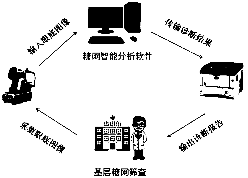
[0034] like figure 1 As shown, in this embodiment, the screening system includes a color fundus camera, image processing and screening equipment (it can be implemented by a computer equipped with sugar network intelligent analysis software, and can also be implemented by a dedicated processor) and Diagnostic report printer. Images collected by other hospitals or primary screening units can also be transmitted to image processing and screening equipment.

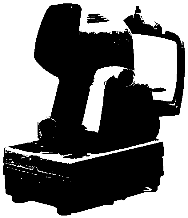
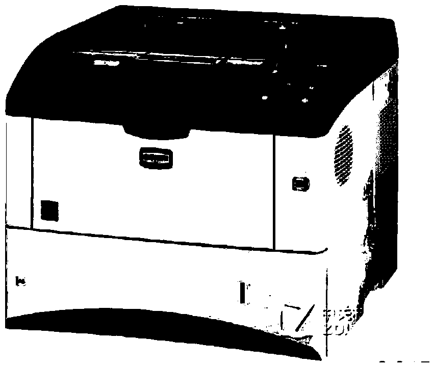
[0035] figure 2 A schematic structural view of the color fundus camera used in this embodiment is shown; image 3 A schematic structural diagram of the diagnostic report printer used in this embodiment is shown. The color fundus camera is used to obtain the patient's color fundus image, which is operated by a professional doctor and transmitted to the fixed path of the computer.

[0036] The collected images are directly or indirectly transmitted to the image processing and screening equipment, which performs image proce...

Embodiment 2

[0051] In this embodiment, the screening system includes a fundus image receiver, a computer (which is installed with sugar net intelligent analysis software) and a diagnostic report display device.

[0052] In this embodiment, the fundus image receiver remotely receives fundus images sent by other non-local fundus cameras. The computer includes a processor and a memory, and sugar net intelligent analysis software is installed in the computer.

[0053] Sugar network intelligent analysis software can execute Figure 4 The individual process steps shown in . As shown in the figure, the fundus image obtained by a remote fundus camera or other equipment is first received, and then image preprocessing is performed.

[0054] Next, image quality correction, blood vessel segmentation, optic disc positioning, red lesion extraction, and brightness lesion extraction are performed. Then, use the characteristic information such as blood vessels, optic discs, and lesions obtained in the ...

Embodiment 3

[0057] In this embodiment, the structure of the system is the same as that in Embodiment 1, except that the optic disc positioning module adopts a special positioning method.

[0058] Specifically, in this embodiment, the optic disc positioning module proposes for the first time a method based on the human visual attention model to locate the optic disc by means of some advances in computational neuroscience. The principle is as follows: First, multiple feature maps of different scales are generated. Specifically, after image preprocessing, through Gaussian filtering and continuous downsampling, Gaussian pyramids of multiple different scales are extracted from the image. "Operation calculation. Generate brightness, color, and orientation feature maps, such as Figure 7 Shown: Among them, (a) is the brightness saliency map, (b) is the color saliency map, (c) is the direction saliency map.

[0059] Based on the 3 feature maps, establish 3 feature saliency descriptions and ...


PUM


Abstract

The invention provides a system for screening diabetic retinopathy. The system comprises an eyeground image obtaining device, an image processing and screening device and a report output device, wherein the eyeground image obtaining device is used for acquiring or receiving eyeground images of detected personnel; the image processing and screening device is used for processing the eyeground images and detecting whether pathology exists and then transmitting a detection result to the report output device; and the report output device outputs a corresponding detection report on the basis of the detection result. According to the system, the sensitivity of determining the diabetic retinopathy can reach 93.8%, the specificity can reach 94.5%, the precision is equivalent to manual determination precision, yet the detection time is greatly reduced, and the average detection time is 8.7 seconds, which is only one thirtieth of manual film reading time. Therefore, the scanning cost of the diabetic retinopathy can be substantially decreased, the diabetic retinopathy can be found early and treated early, the pain of patients is greatly reduced, and the clinical application prospect and the social benefits are quite good.

Description

technical field [0001] The invention relates to the field of medical equipment, in particular to a system for screening diabetic retinopathy. Background technique [0002] Diabetic retinopathy, referred to as sugar retinopathy (DR), is one of the most common chronic complications of diabetes. According to the World Health Organization (WHO), by 2030, the number of DR patients in the world will increase to 366 million, and it has become one of the four major blindness diseases. The prevention and treatment of DR will become a serious worldwide problem. [0003] Studies have shown that early diagnosis and treatment of DR patients can effectively prevent visual loss and blindness, and the key to prevention is through fundus photographic examination, regular follow-up to find the progress of the disease, and timely laser intervention. However, more than 50% of diabetic patients in the world have not received any form of eye examination, and the screening of diabetic retinopathy...

Claims


Application Information

Patent Timeline
Patent Type & Authority Applications(China)
IPC IPC(8): G06T7/00
CPCG06T7/0012G06T2207/30041
Inventor 朱江兵柯鑫王茜李建军何建国潘津陈绍义王亚鹏
Owner BEIJING DAHENG IMAGE VISION
Why Eureka
Try Eureka