Method for the analysis of N-glycans attached to immunoglobulin G from human blood plasma and its use

一种免疫球蛋白、分析方法的技术,应用在人类血浆中的免疫球蛋白G连接的N-聚糖的分析及其应用领域

Inactive Publication Date: 2016-05-11
ZHONGKE YAMEI MEDICAL TECH (BEIJING) CO LTD
View PDF3 Cites 6 Cited by
  • Summary
  • Abstract
  • Description
  • Claims
  • Application Information

AI Technical Summary

Problems solved by technology

Because of the complexity of the aging process, no single parameter can determine the biological age of an individual

Method used


Image

  • Method for the analysis of N-glycans attached to immunoglobulin G from human blood plasma and its use
  • Method for the analysis of N-glycans attached to immunoglobulin G from human blood plasma and its use
  • Method for the analysis of N-glycans attached to immunoglobulin G from human blood plasma and its use

Examples


Embodiment 1

[0179] Example 1 Purification of Immunoglobulin G (IgG) from Plasma

[0180] Immunoglobulin G (IgG) was purified from plasma by affinity chromatography with the aid of protein G bound to a monolithic column in a 96-well microplate (BiaSeparations, Ljubljana, Slovenia). All solutions used in the purification process were prepared with ultrapure water and filtered through 0.2 PES filters. All washing steps through the monolith were accomplished with the aid of a vacuum pump (Pall Corporation, MI, USA). Before use, the chromatographic column was washed with 2 ml of water and activated with 4 ml of binding buffer (lxPBS; pH=7.4). Prior to loading on the monolithic column, 100 ml of plasma was diluted with binding buffer (1:7, V / V) and filtered through a 0.45 μm pore size GHP filter plate. After dilution and loading of filtered plasma, the monolithic chromatography plate was washed 3 times with 2 mL of binding buffer to remove unbound protein. The IgG antibody was eluted fro...

Embodiment 2

[0181] Example 2 Release of Glycans from Immunoglobulin G (IgG)

[0182] An aliquot of purified IgG (app. 100 μg) was dried in a vacuum centrifuge, then resuspended and denatured in 30 μl of 1.33% SDS solution at 65° C. for 10 minutes. Subsequently, 10 μl of 4% NP40, 10 μl of 5-fold PBS and 1.25 mUP PNGazeF (N-glycosidase F, ProZyme, California, USA) were added. The mixture was incubated overnight at 37°C.

Embodiment 3

[0183] Example 3 Labeling IgG Glycans and Removing Excess Fluorescent Labels

[0184] After deglycosylation, add 25 μl of fluorescently labeled mixture to the reaction mixture: a mixture of acetic acid and dimethyl sulfoxide containing 0.48 mg of 2-aminobenzamide (2AB) and 1.12 mg of 2-picoline borane ( 30%:70%, V / V). The mixture was incubated at 65°C for 2 hours. Excess markers were removed by means of microcrystalline cellulose added to 0.45 μm pore size GHP filter plates. Add 200 μl of 0.1 g / mL cellulose aqueous suspension to each well of the filter plate. The cellulose was washed 5 times with 200 μl of water and 3 times with 80% acetonitrile for activation. After labeling, the reaction mixture diluted with 400 μl of acetonitrile was added to the cellulose. Wash the cellulose 7 times with 200 μl of 80% acetonitrile to remove excess marker. Wash the cellulose twice with 100 μl of water to elute the labeled immunoglobulin glycans. The eluate of IgG glycan 2-aminoben...


PUM


Abstract

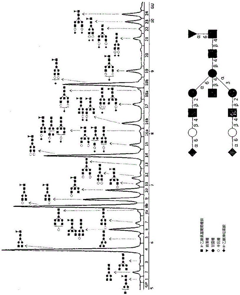
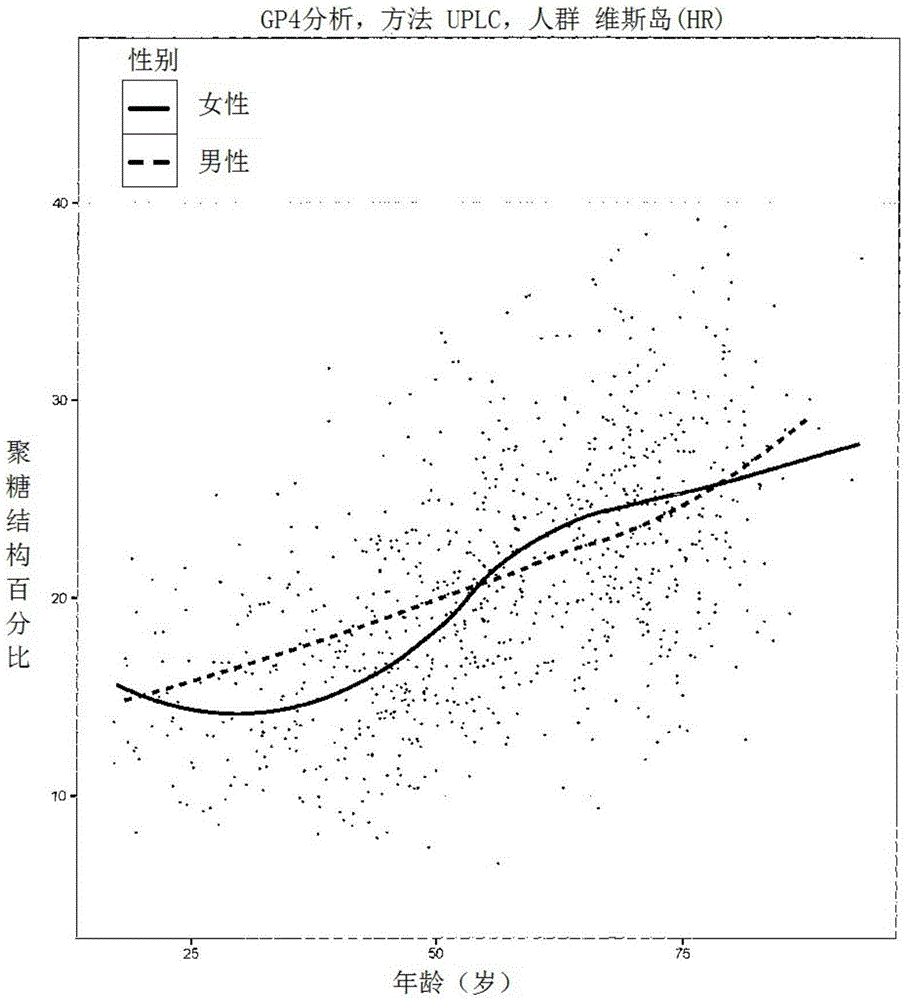
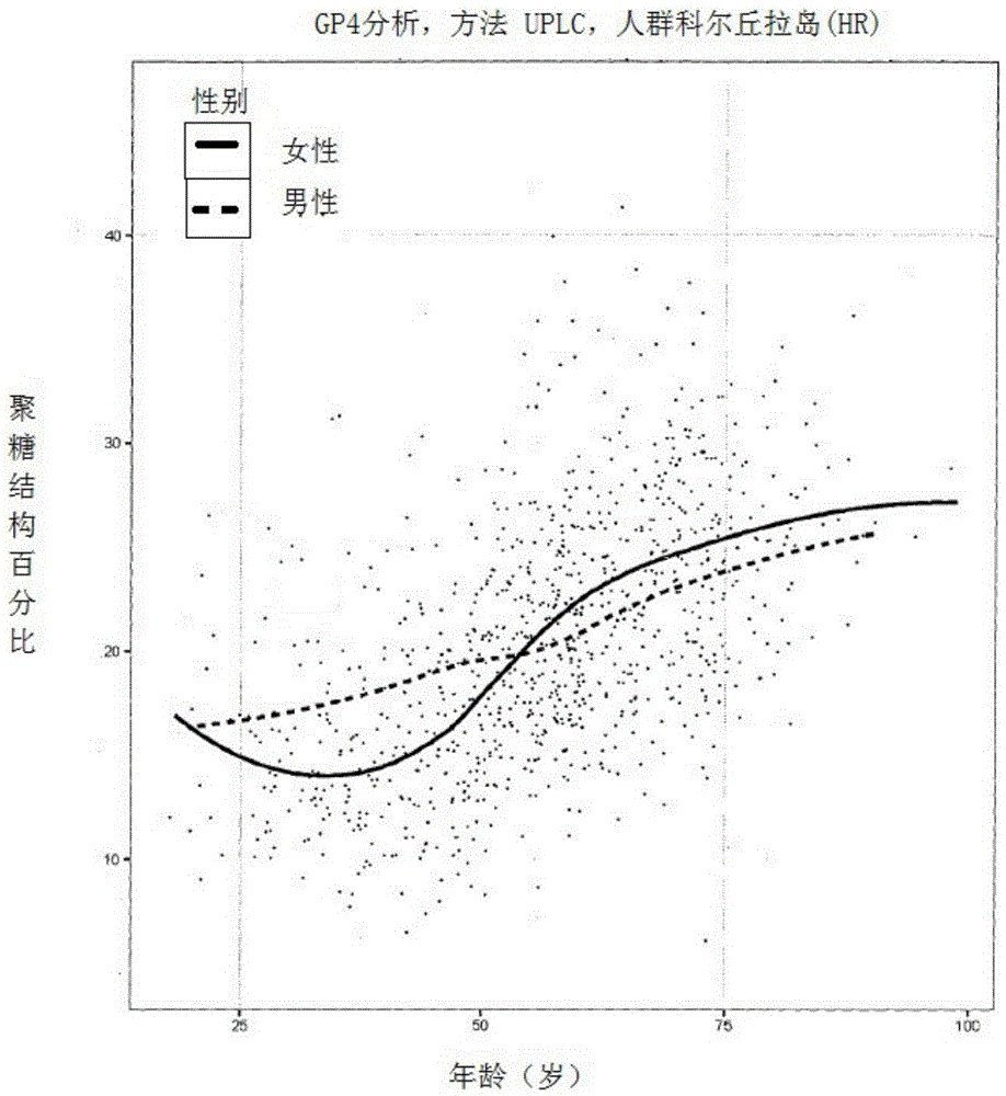
The invention discloses a method for the analysis of N-glycans attached to immunoglobulin G (IgG) or IgG N-glycopeptides from human blood plasma in which relative abundance of two or more glycans is determined, out of total six, and for these glycans it is determined that they strongly correlate with age. The glycans have the following structures: F(6)A2 (GP4): R1, R2, R3, R4=H F(6)A2B (GP6): R1=GlcNAc; R2, R3, R4 H F(6)A2[6]G1 (GP8): R1, R3, R4=H; R2= Gal F(6)A2G2 (GP14): R1=H; R2, R3=Gal; R4=H F(6)A2BG2 (GP15): R1=GlcNAc; R2, R3=Gal; R4=H F(6)A2G2S1 (GP18): R1=H; R2, R3=Gal; R4=NeuAc GlcNAc=N-acetylglucosamine Fuc=fucose Man=mannose NeuAc=N-acetylneuraminic acid Gal=galactose From the results of the analysis, Glycan Age Index (GAI) is calculated, and it is useful for: prediction of biological age of a tested individual; monitoring efficacy of methods that slow down the ageing process; monitoring progression of diseases that are developed as a result of the ageing process advancement, like: inflammatory diseases (including atherosclerosis), autoimmune diseases, tumours, diabetes, arthritis, osteoporosis, and Alzheimer disease; and evaluation of overall condition / health of a body.

Description

technical field [0001] The present invention relates to a method for the analysis of immunoglobulin G-linked N-glycans in human plasma, the purpose of which is to: more accurately predict the biological and / or chronological age of a person; possibly be used to monitor the effect of a method for delaying the aging process; May be used to monitor the progression of diseases resulting from accelerated aging processes such as: inflammatory diseases (including atherosclerosis), autoimmune diseases, tumors, diabetes, arthritis, osteoporosis and Alzheimer's disease; and Assess the general condition / health of a person. [0002] technical problem [0003] The invention solves the technical problem of accurate prediction of human biological age by quantitatively analyzing two or more immunoglobulin G (IgG)-linked N-glycans (6 in total) most closely related to age. [0004] In addition, the present invention solves the following problems, which may be used to effectively monitor the ef...

Claims


Application Information

Patent Timeline
Patent Type & Authority Applications(China)
IPC IPC(8): G01N33/53G01N33/68
CPCG01N33/5308G01N33/6851G01N2400/10G01N30/72G01N33/6854G01N2030/027G01N2333/976G01N2440/38G01N2800/52G01N2800/7042
Inventor 戈丹·劳克玛雅·普希奇宫巴克维奇福拉诺·黄片
Owner ZHONGKE YAMEI MEDICAL TECH (BEIJING) CO LTD
Why Eureka
Try Eureka