Profile model for simulating performance of tumor contrast-enhanced ultrasonography and applied in tumor diagnosis and treatment study

A technology of contrast-enhanced ultrasound and ultrasound contrast agent, which is applied in the medical field and can solve the problems of distinguishing and unusable normal tissue models

Active Publication Date: 2016-05-18
THE THIRD AFFILIATED HOSPITAL OF SUN YAT SEN UNIV
View PDF8 Cites 6 Cited by
  • Summary
  • Abstract
  • Description
  • Claims
  • Application Information

AI Technical Summary

Problems solved by technology

[0005] However, the above-mentioned phantoms are all models based on two-dimensional ultrasound imaging. Although the tumor model is hyperechoic in two-dimensional ultrasound imaging, it is still anechoic in harmonic contrast ultrasound imaging mode and cannot be compared with normal tissue. Model distinction, cannot be used for contrast-enhanced ultrasound and ultrasound-guided tumor biopsy or ablation training based on contrast-enhanced ultrasound

Method used


Image

  • Profile model for simulating performance of tumor contrast-enhanced ultrasonography and applied in tumor diagnosis and treatment study
  • Profile model for simulating performance of tumor contrast-enhanced ultrasonography and applied in tumor diagnosis and treatment study
  • Profile model for simulating performance of tumor contrast-enhanced ultrasonography and applied in tumor diagnosis and treatment study

Examples


Embodiment 1

[0084] Embodiment 1 Preparation of Nano Lipid Microbubble Ultrasonic Contrast Agent

[0085] 1. Dissolve the phospholipid components dipalmitoyl phosphatidylcholine, distearoylphosphatidylethanolamine and dipalmitoylphosphatidic acid in chloroform. The mass-number ratio of dipalmitoylphosphatidylcholine, distearoylphosphatidylethanolamine and dipalmitoylphosphatidic acid is 18:1:3.5.

[0086] 2. Add double distilled water containing 10% glycerol (v / v) and 2mg / mL Pluronic F68 after the chloroform volatilizes naturally to form a phospholipid film, and hydrate at 37°C for 1 hour. Acoustic vibration was performed at the liquid-liquid junction for 5 minutes, and octafluoropropane gas was introduced at the same time.

[0087] 3. Centrifuge the microbubble solution at 50×g for 5 minutes, absorb the milky white liquid in the lower layer and transfer it to a tube, centrifuge at 805×g for 30 minutes, discard the lower layer liquid, and resuspend it with PBS to obtain nano-lipid microbu...

Embodiment 2

[0089] The preparation of embodiment 2 phantom model

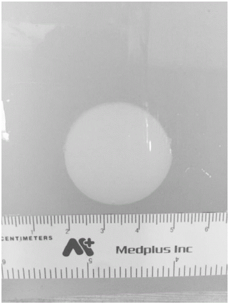
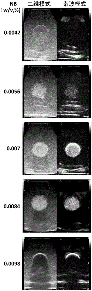
[0090] Weigh 20 g of carrageenan and 1 g of gastric window acoustic contrast agent in a beaker with an electronic balance, and add 1000 mL of double distilled water at 60°C. Place the beaker in a constant temperature water bath at 60°C and stir with a glass rod until the solute is completely dissolved to form normal tissue matrigel. Take 38 mL of Matrigel in a small beaker, add 2 mL of milk to form tumor gel Matrigel, then add nano-lipid microbubble ultrasound contrast agent, stir evenly to form tumor gel. In this example, tumor gel models with nanoscale lipid microbubble ultrasound contrast agent concentrations (calculated by phospholipid mass) of 0.0042%, 0.0056%, 0.007%, 0.0084% and 0.0098% (w / v) were prepared respectively. After placing the tumor glue in a sphere mold with a diameter of 3 cm, place it in ice water and cool it for 5 minutes, and take it out to obtain the tumor glue model.

[0091] The tumor gel model ...

Embodiment 3

[0092] Embodiment 3 Ultrasonic imaging effect detection of phantom model

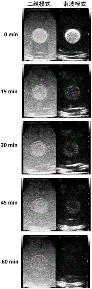
[0093]The phantom phantoms with different concentrations of nanoscale contrast agents were examined with contrast-enhanced ultrasound. In this embodiment, Esaote MyLabtwice model clinical ultrasonic diagnostic instrument and linear array probe are used to detect the phantom, the imaging frequency band is 3-11 MHz, the focal point is placed in the middle of the tumor, and the gain is 0.5. For harmonic contrast ultrasound imaging mode, use a mechanical index of 0.03 and a gain of 0.38. 10 min after the phantom model was taken out of the refrigerator was defined as 0 min, and two-dimensional ultrasound and harmonic contrast ultrasound imaging were performed on the phantom model at different time points.

[0094] The result is as figure 2 As shown, at the time point of 0 min, phantom models prepared with different concentrations of nano-scale contrast agents showed different imaging effects. When the co...


PUM


Abstract

The invention belongs to the field of medicine and relates to a profile model for simulating the performance of the tumor contrast-enhanced ultrasonography and applied in the tumor diagnosis and treatment study. The profile model is characterized by comprising a tumor gel used for simulating a tumor, and a normal tissue gel used for simulating the surrounding normal tissue. The tumor gel comprises a nano micro-bubble ultrasonic contrast agent. The nano micro-bubble ultrasonic contrast agent is distributed uniformly, good in dispersibility, strong in imaging effect in vivo and in vitro, and better in stability in the high/low-frequency ultrasonic sound field. Therefore, the profile model facilitates the contrast-enhanced ultrasonic scanning process of a user, the practice of the tumor aspiration biopsy and the practice of the tumor ablation therapy with the help of the high/low-frequency ultrasonic guidance.

Description

technical field [0001] The invention belongs to the field of medicine, and relates to a phantom model for simulating tumor ultrasound contrast imaging. Specifically, it relates to the field of ultrasound-enhanced ultrasound and ultrasound-guided liver cancer diagnosis and interventional therapy in ultrasound medicine, and relates to an improved model for teaching and training of ultrasound-enhanced ultrasound technology, high / low frequency ultrasound-guided puncture biopsy technology and tumor ablation technology . technical background [0002] The contrast-enhanced ultrasound technology is considered to be the third revolution in the development of ultrasound medicine, which advances the diagnostic mode of ultrasound from conventional morphological diagnosis to functional diagnosis. In 2013, after in-depth analysis of a large number of prospective clinical studies, Westwood et al. proposed that the diagnostic efficiency of contrast-enhanced ultrasound technology for focal ...

Claims


Application Information

Patent Timeline
Patent Type & Authority Applications(China)
IPC IPC(8): G09B23/28
CPCG09B23/286
Inventor 周慧超尹庭辉许尔蛟李凯郑荣琴吕淑敏
Owner THE THIRD AFFILIATED HOSPITAL OF SUN YAT SEN UNIV
Why Eureka
Try Eureka