A magnetic resonance imaging method and device for deep vein thrombosis of lower extremities

A deep vein thrombosis and magnetic resonance imaging technology, applied in the field of medical imaging, can solve problems such as dependence, blood flow signal contamination of imaging results, and inability to use pregnant women.

Active Publication Date: 2019-08-02
SHENZHEN INST OF ADVANCED TECH +1
View PDF7 Cites 0 Cited by
  • Summary
  • Abstract
  • Description
  • Claims
  • Application Information

AI Technical Summary

Problems solved by technology

[0003] Magnetic resonance imaging techniques commonly used in the clinical diagnosis of DVT mainly include Contrast Enhanced Magnetic Resonance Venography (CE-MRV), balanced steady-state free precession imaging (balanced Steady-state Free Precession, bSSFP) and There are three types of Magnetic Resonance Direct Thrombus Imaging (MRDTI) based on inversion gradient echo, among which CE-MRV is often used to determine the presence or absence of thrombus and the distribution range, and can be based on the enhancement of thrombus and vessel wall However, the CE-MRV examination needs to accurately estimate the circulation time of the target blood vessel, and the scanning operation is difficult. At the same time, the use of contrast agent not only increases the examination cost, but also cannot be used for severe renal function. Incomplete and pregnant patients, and the natural bright blood effect of bSSFP technology makes the blood flow in the lumen of the vessel show high signal and the thrombus shows low signal without the use of contrast agent. MRV is similar, but bSSFP technology can only be used for rapid screening of DVT, the size of thrombus is not clearly displayed, and the age of thrombus cannot be judged. It is also susceptible to banding artifacts caused by the inhomogeneity of the main magnetic field of the magnetic resonance system. As for the technique of MRDTI, it is a T1-weighted gradient echo sequence, which uses the unique paramagnetic properties of methemoglobin, which exists in large quantities in subacute thrombus, to directly image the thrombus, and has high specificity and sensitivity. However, the MRDTI technique relies too much on the content of methemoglobin to shorten the T1 relaxation time, so that the thrombus appears as a bright signal in the image, so it can only diagnose thrombus with a high content of methemoglobin (acute→subacute) or subacute phase, Ineffective against hyperacute and chronic thrombus with low methemoglobin content
Recently, a magnetic resonance vessel wall imaging technique was proposed to achieve clinical examination and diagnosis of DVT, but this technique uses a three-dimensional fast spin-echo sequence, which has very limited suppression effect on blood flow signals and cannot completely suppress venous vessels. The blood flow signal in the cavity makes the imaging result easily polluted by the blood flow signal, and it is difficult to accurately distinguish the vessel wall, blood flow and thrombus

Method used


Image

  • A magnetic resonance imaging method and device for deep vein thrombosis of lower extremities
  • A magnetic resonance imaging method and device for deep vein thrombosis of lower extremities
  • A magnetic resonance imaging method and device for deep vein thrombosis of lower extremities

Examples


Embodiment Construction

[0026] In order to make the object, technical solution and beneficial effects of the present invention more clear, the present invention will be further described in detail below in conjunction with the accompanying drawings and embodiments. It should be understood that the specific embodiments described here are only used to explain the present invention, not to limit the present invention.

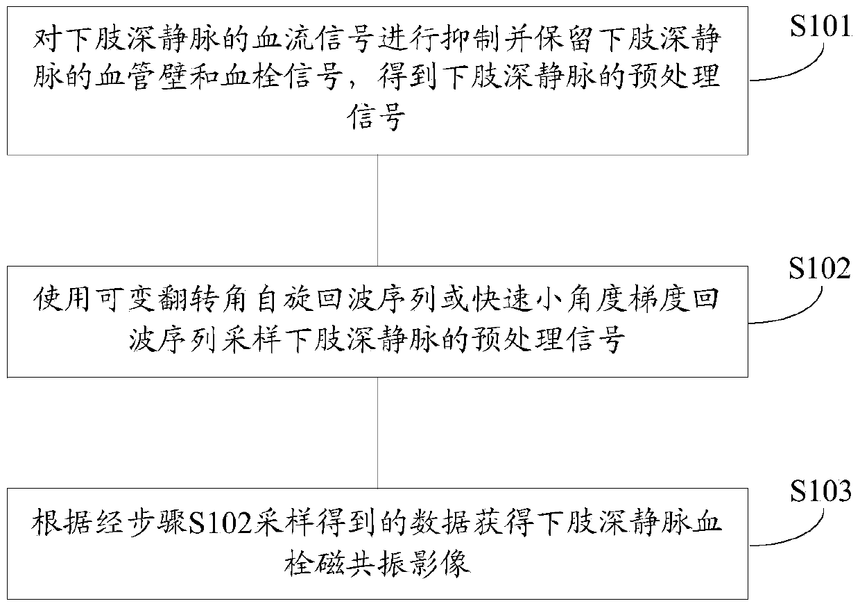
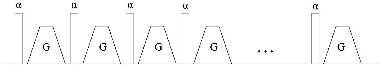
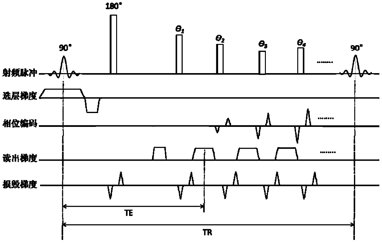
[0027] An embodiment of the present invention provides a magnetic resonance imaging method for deep vein thrombosis of lower extremities, the method comprising: suppressing the blood flow signal of the deep vein of the lower extremity and retaining the vessel wall and thrombus signals of the deep vein of the lower extremity to obtain the deep vein of the lower extremity Preprocessing the signal; sampling the preprocessing signal of the deep vein of the lower extremity by using a variable flip angle spin echo sequence or a fast small-angle gradient echo sequence; obtaining a magnetic reson...


PUM


Abstract

The invention provides a lower limb deep venous thrombus magnetic resonance imaging method and device. On the premise that no contrast media are used, injuries to a patient are avoided, and meanwhile the imaging effect is improved. The method includes the steps that blood flow signals of lower limb deep veins are suppressed, blood vessel wall and thrombus signals of the lower limb deep veins are retained, and preprocessed signals of the lower limb deep veins are obtained; the preprocessed signals of the lower limb deep veins are sampled through a variable-turning-angle spin echo sequence or a fast small-angle gradient echo sequence; according to sampled data, a lower limb deep venous thrombus magnetic resonance image is acquired. By means of the technical scheme, on one hand, blood vessel walls, blood flow and thrombi of the lower limb deep veins can be accurately differentiated, and whether thrombi exist in blood vessels or not can be visually displayed; on the other hand, the lower limb deep venous thrombus magnetic resonance image obtained through the method and device can display whether thrombi exist in the lower limb deep veins or not and the distribution positions of the thrombi, the thrombi can be preliminarily staged, and practical and effective information is provided for clinical treatment.

Description

technical field [0001] The invention belongs to the field of medical imaging, in particular to a magnetic resonance imaging method and device for deep vein thrombosis of lower extremities. Background technique [0002] At present, there are four types of imaging techniques that can be used in the examination of deep vein thrombosis (DVT) of the lower extremities, including venous angiography (Digital Subtraction Angiography, DSA), ultrasound, CT, and magnetic resonance imaging. These four types of methods have their own advantages and disadvantages. For example, DSA can accurately determine the presence or absence of thrombus and its distribution range, but this method is invasive and only used in the process of interventional treatment; although ultrasound is noninvasive, it is convenient and simple. However, it relies too much on the doctor's technique and experience, and this technology can only determine the distribution range of thrombus, but cannot determine the age of...

Claims


Application Information

Patent Timeline
Patent Type & Authority Patents(China)
IPC IPC(8): A61B5/055
CPCA61B5/055A61B5/7225
Inventor 陈汉威谢国喜梁健科刘新任亚楠叶裕丰邓炜
Owner SHENZHEN INST OF ADVANCED TECH
Why Eureka
Try Eureka