Imaging model in small animal living bodies with echinococcus granulosus and construction method thereof

A technology of echinococcus small animals and imaging models, which is applied in the field of bioengineering, can solve the problems of blank imaging models, and achieve the effect of maintaining the survival of cells and maintaining energy balance

Inactive Publication Date: 2016-06-08
王思博
View PDF2 Cites 10 Cited by
  • Summary
  • Abstract
  • Description
  • Claims
  • Application Information

AI Technical Summary

Problems solved by technology

[0004] The purpose of the present invention is to provide a small animal Echinococcus granulosus in vivo imaging model and its construction method, aiming to solve the problem that the Echinococcus granulosus small animal in vivo imaging model is still blank in the biological field

Method used


Image

  • Imaging model in small animal living bodies with echinococcus granulosus and construction method thereof
  • Imaging model in small animal living bodies with echinococcus granulosus and construction method thereof
  • Imaging model in small animal living bodies with echinococcus granulosus and construction method thereof

Examples


Embodiment Construction

[0029] In order to make the objectives, technical solutions and advantages of the present invention clearer, the present invention will be further described in detail below with reference to the embodiments. It should be understood that the specific embodiments described herein are only used to explain the present invention, but not to limit the present invention.

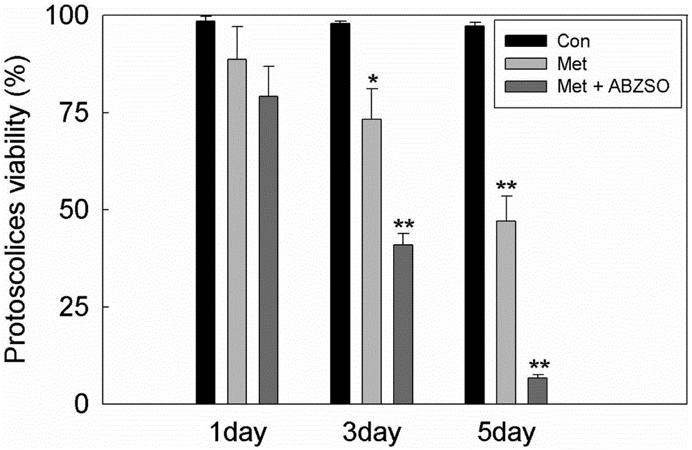
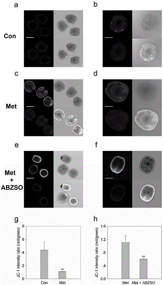
[0030] The invention constructs a fluorescence imaging model in mice; through this model, the activity of protoscoria can not only be analyzed by using the detected fluorescence intensity and the ratio of red-green fluorescence, but also helps to establish a long-term stable luminescent mouse living body in the future Model.

[0031] The application principle of the present invention will be described in detail below with reference to the accompanying drawings.

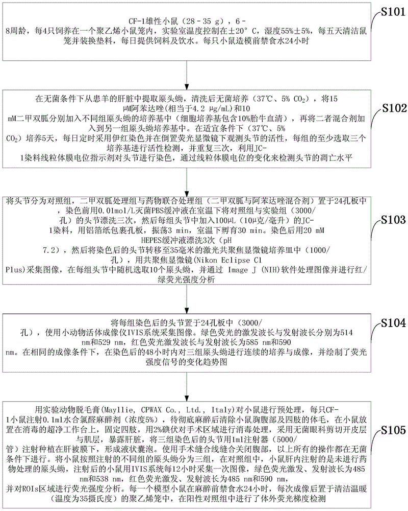
[0032] like figure 1 As shown, the construction method of the small animal in vivo imaging model of Echinococcus granulosus according to the embodiment...


PUM


Abstract

The invention discloses an imaging model in small animal living bodies with echinococcus granulosus and a construction method thereof. The construction method comprises the steps of: feeding CF-1 male mice; carrying out in vitro culture and drug treatment on protoscolex; carrying out in vitro fluorescence imaging and fluorescence gradient analysis; pretreating mice with depilatory paste for experimental animals, injecting the abdominal cavity of each mouse with chloral hydrate anesthetic, implanting three groups of stained scoleces under Glisson capsules by injection, dividing the mice into three groups according to different groups of injected protoscolex; in a control group, injecting the livers of mice with protoscolex on which drug treatment is not carried out, collecting images every 12 hours after injection, carrying out fluorescence intensity analysis on the ROIs region, putting into a polyethylene cage after imaging, and carrying out in vitro fluorescence gradient detection in the positive control group. The construction method constructs a mouse model with echinococcus granulosus which can express luciferase, provides a platform for drug sensitive tests in living bodies with echinococcosis, and can monitor the growth and transfer of hydatid in the living model dynamically in a long term.

Description

technical field [0001] The invention belongs to the technical field of bioengineering, and in particular relates to a small animal in vivo imaging model of Echinococcus granulosa and a construction method thereof. Background technique [0002] Echinococcus granulosus is a common zoonotic disease that not only harms public health but also affects economic development. Dogs and foxes are the ultimate hosts of infection. Dogs ingest vesicles containing protosegmentum, and humans become infected through contact with dogs, or by mistakenly ingesting water and vegetables containing worm eggs. Infection with Echinococcus granulosus can cause damage to single or multiple organs, the parasitic organs mainly include liver (70%), lung (20%), and 10% may be parasitic in any part of the body (such as brain and muscle tissue) ). The geographic distribution of hydatid disease is wide, mainly concentrated in parts of Eurasia, including the Mediterranean region, Russia, Central Asia, the c...

Claims


Application Information

Patent Timeline
Patent Type & Authority Applications(China)
IPC IPC(8): A61K49/00A61K35/62A01K67/027
CPCA61K49/0008A01K67/027A01K2207/20A01K2227/105A01K2267/03A61K35/62A61K49/0019A61K49/006
Inventor 吴向未王思博陈雪玲杨涛夏杰
Owner 王思博
Why Eureka
Try Eureka