Attenuation correction method based on image segmentation

A technology of attenuation correction and image segmentation, applied in the field of nuclear medicine imaging, can solve the problems of manual intervention, increase of doctor's workload, errors, etc., achieve automatic detection, improve image reconstruction speed, and reduce human errors.

Active Publication Date: 2016-06-15
INST OF HIGH ENERGY PHYSICS CHINESE ACADEMY OF SCI
View PDF4 Cites 6 Cited by
  • Summary
  • Abstract
  • Description
  • Claims
  • Application Information

AI Technical Summary

Problems solved by technology

At present, in the attenuation correction of head-specific functional imaging equipment, this method mainly draws the contour of the head manually or semi-automatically. The disadvantage is that manual in

Method used


Image

  • Attenuation correction method based on image segmentation
  • Attenuation correction method based on image segmentation
  • Attenuation correction method based on image segmentation

Examples


Embodiment Construction

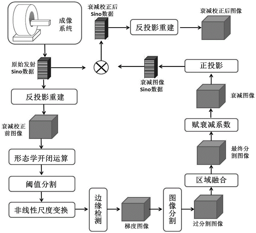
[0026] The technical solutions in the embodiments of the present invention are clearly and completely described below in conjunction with the drawings in the embodiments of the present invention. Apparently, the described embodiments are only a part of the present invention, not all of them. Based on the embodiments of the present invention, all other embodiments obtained by persons of ordinary skill in the art without making creative efforts belong to the protection scope of the present invention. The method of the present invention comprises:

[0027] 1. Reconstruct the original emission data to obtain the initial image model (there are many data reconstruction algorithms at present, the so-called data reconstruction refers to reconstructing the scanned data through a specific algorithm to obtain the scanned image). The initial image model will be used for image segmentation processing, so it should be as smooth as possible. This requires that attention should be paid to t...


PUM


Abstract

The invention discloses an attenuation correction method based on image segmentation. The method comprises the following steps: 1) carrying out reconstruction on original emission data of a nuclear imaging system; 2) carrying out filtering and de-noising processing on the obtained reconstructed image, and then, carrying out threshold segmentation, nonlinear scale transformation and differential treatment to obtain a gradient image; 3) carrying out segmentation on the gradient image according to the gradient distribution of the gradient image; 4) carrying out region merging on the segmented gradient images to obtain final segmented images; 5) providing a corresponding linear attenuation coefficient for each segmented image to obtain corresponding attenuation images; 6) carrying out orthographic projection on each attenuation image and carrying out correction on the original emission data according to the projection data; and 7) carrying out reconstruction on the corrected original emission data to obtain an image after attenuation correction. Compared with the prior art, the method does not need manual intervention, and can reduce personal errors. Besides, the method is quick in realization speed and can improve image reconstruction speed of the system.

Description

technical field [0001] The invention belongs to the field of nuclear medicine imaging and relates to an attenuation correction method based on image segmentation. Background technique [0002] Functional nuclear medicine imaging technology (Positron Emission Tomography (PET), Single Photon Emission Tomography (Single Photon Emission Tomography)) as an advanced imaging technology represents the highest level of nuclear medicine. In recent years, the application of functional nuclear imaging systems in the field of cranial nerve research has attracted more and more attention in the clinical and scientific research fields. At the same time, high-resolution 3D imaging systems and corresponding image reconstruction and correction algorithms have also made great progress. However, the current imaging level cannot meet the requirements of brain function research for image quality and quantitative accuracy, which requires continuous exploration and improvement of imaging system desi...

Claims


Application Information

Patent Timeline
IPC IPC(8): G06T5/00G06T5/10G06T7/00G06T15/10G06T11/00
CPCG06T5/002G06T5/10G06T11/008G06T15/10G06T2207/10104G06T2207/10108G06T2207/30016G06T2215/06
Inventor 马波柴培章志明魏龙刘双全李默涵朱锦霞顾笑悦李道武
Owner INST OF HIGH ENERGY PHYSICS CHINESE ACADEMY OF SCI
Why Eureka
Try Eureka