
A Blood Vessel Segmentation Method for Liver CTA Sequence Images

A technology of liver blood vessels and sequence images, applied in the field of medical image processing, can solve the problems of ineffective segmentation of small blood vessels, low contrast, and ineffective extraction.

Active Publication Date: 2018-09-21
湖南提奥医疗科技有限公司
View PDF4 Cites 0 Cited by
  • Summary
  • Abstract
  • Description
  • Claims
  • Application Information

AI Technical Summary

Problems solved by technology

Single gray-scale or gradient-based segmentation methods, such as 3D region growing, fuzzy clustering, etc., cannot effectively extract low-contrast liver vessels
In recent years, the active contour model and its hybrid model have been widely used in 3D blood vessel segmentation, but the evolution surface of this type of model is easy to cross the weak boundary of the blood vessel, resulting in serious over-segmentation, and the initial area of ​​the blood vessel needs to be provided interactively
In addition, the above methods are difficult to segment small blood vessels

Method used


Image

  • A Blood Vessel Segmentation Method for Liver CTA Sequence Images
  • A Blood Vessel Segmentation Method for Liver CTA Sequence Images
  • A Blood Vessel Segmentation Method for Liver CTA Sequence Images

Examples


Embodiment Construction

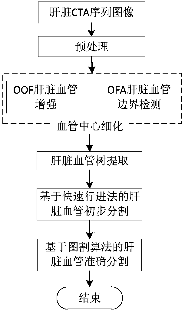
[0072] figure 1 It is a flow chart of the blood vessel segmentation method for liver CTA sequence images implemented in the present invention. Firstly, the window width / window level is adjusted from the input liver blood vessel image, the contrast of blood vessels is improved, and the noise is smoothed by anisotropic filtering. Then, the OOF and OFA methods are used to enhance the vessels and their boundaries, and optimize the central response of the vessels. Next, according to the geometric structure of the vessel, the centerline of the vessel is extracted and a vessel tree is constructed. Finally, the fast marching method is used to initially segment the liver blood vessels, and the graph cut energy function is constructed by combining the gray distribution of the initial blood vessels and the background, and the energy function is optimized to achieve accurate segmentation of liver blood vessels.

[0073] Combine below figure 1 , using an embodiment to describe in detail...


PUM


Abstract

The invention discloses a blood vessel segmentation method for liver CTA sequence images. First, contrast enhancement and smooth noise preprocessing are performed on the input three-dimensional liver sequence image. Then, OOF and OFA algorithms are used to enhance liver blood vessels and their boundaries, and refine the blood vessel center; according to According to the geometric structure of blood vessels, the seed points of the blood vessel centerline are automatically searched, the centerline of the liver blood vessels is extracted, and the liver blood vessel tree is constructed; finally, the liver blood vessels are preliminarily segmented using the fast marching method and the corresponding blood vessel and background grayscale histograms are calculated, using Graph cut algorithm achieves accurate segmentation of liver blood vessels. For CTA sequence images with low contrast, strong noise and blurred boundaries, the present invention makes full use of the geometric shape and grayscale information of blood vessels, and can effectively and accurately segment liver blood vessels. The blood vessel segmentation method of liver CTA sequence images of the present invention can be extended to other three-dimensional blood vessel segmentation.

Description

technical field [0001] The invention belongs to the field of medical image processing, and relates to liver blood vessel enhancement, central line extraction and liver blood vessel segmentation in CTA sequence images. Background technique [0002] Liver vascular segmentation and 3D reconstruction help to accurately obtain the overall information of abdominal liver vascular tissue, which is the premise of computer-aided liver disease diagnosis and liver surgery planning. CTA (computed tomography angiography) is a non-invasive imaging technique with advantages of high density resolution and less damage to the human body, and is widely used in the assessment and diagnosis of liver diseases. Due to the complex structure of liver blood vessels, the intertwining of blood vessels, and the large differences between different individuals, the segmentation of liver blood vessels is facing great challenges. In clinical applications, in order to construct a liver vascular model, radiol...

Claims


Application Information

Patent Timeline
Patent Type & Authority Patents(China)
IPC IPC(8): G06T5/00G06T7/11
CPCG06T7/0012G06T2207/30101G06T5/70
Inventor 赵于前曾业战廖苗杨勍杨少迪
Owner 湖南提奥医疗科技有限公司
Learn More