Image visualization method for detecting single molecule DNA duplication

A single-molecule, single-stranded technology, applied in the field of image visualization to detect single-molecule DNA replication, which can solve problems such as unsolvable description and characterization, lack of direct observation methods, etc.

Inactive Publication Date: 2016-07-13
SHANGHAI INST OF APPLIED PHYSICS - CHINESE ACAD OF SCI
View PDF3 Cites 5 Cited by
  • Summary
  • Abstract
  • Description
  • Claims
  • Application Information

AI Technical Summary

Problems solved by technology

However, in terms of image visualization, at the single-molecule level, there are currently only reports on image visualization methods for RNA transcription, and for DNA replication at the single-mol

Method used


Image

  • Image visualization method for detecting single molecule DNA duplication
  • Image visualization method for detecting single molecule DNA duplication
  • Image visualization method for detecting single molecule DNA duplication

Examples


Embodiment 1

[0038] The preparation of embodiment 1 single-stranded DNA-DNA origami

[0039] The hollow DNA origami designed in this example utilizes M13mp18DNA and staple single-strand self-assembly to form an equilateral triangle pattern. The preparation method of the equilateral triangle hollow DNA origami is as follows: the ring-shaped long scaffold single-stranded DNA M13mp18 and the two strands A17 and C33 are respectively The staple strand DNA collection ABCL of the sequence shown in SEQIDNO.1 and SEQIDNO.2 is replaced by the sequence table and mixed in TAE-Mg 2+ Buffer system (40mM Tris-acetic acid, 1mM EDTA, 12.5mM MgCl 2 , pH8.0), and then placed in a PCR instrument to anneal from 95°C to 20°C at an annealing rate of 0.1°C / 10s, wherein the two strands of M13mp18, A17 and C33 were replaced by the sequences listed in SEQ ID NO.1 and SEQ ID NO.2, respectively. The molar concentration ratio of the staple strand DNA collection ABCL of the sequence shown is 1:10, with TAE-Mg 2+ The b...

Embodiment 2

[0041] The preparation of embodiment 2 single-stranded DNA-DNA origami

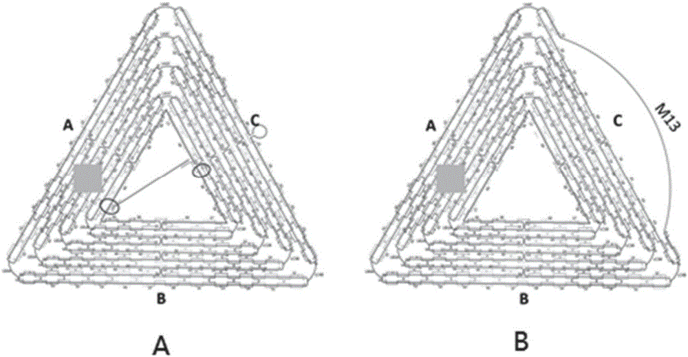
[0042] The hollow DNA origami designed in this example uses M13mp18DNA and the staple chain DNA set ABCL to self-assemble to form an equilateral triangle pattern, wherein the staple chain part of the C side of the equilateral triangle is shortened, specifically, C08, C12, C16, C20, C23, C24, C26, C28, C30, C41, C45, C49, C53, C56, C59, C61, C63 and C64 were replaced by sequences such as sequence listing SEQIDNO.4, SEQIDNO.5, SEQIDNO. 6. SEQ ID NO.7, SEQ ID NO.8, SEQ ID NO.9, SEQ ID NO.10, SEQ ID NO.11, SEQ ID NO.12, SEQ ID NO.13, SEQ ID NO.14, SEQ ID NO.15, SEQ ID NO.16, SEQ ID NO.17, SEQ ID NO.18, The sequences shown in SEQIDNO.19, SEQIDNO.20 and SEQIDNO.21 make the corresponding part of the M13mp18 single-stranded DNA unpaired, forming a DNA strand on the outer edge to become a single-stranded DNA. The components and methods of other assembly systems are the same as in Example 1 , the structure diagram...

Embodiment 3

[0043] Example 3 Real-time dynamic image visualization detection and analysis of DNA replication by atomic force microscope

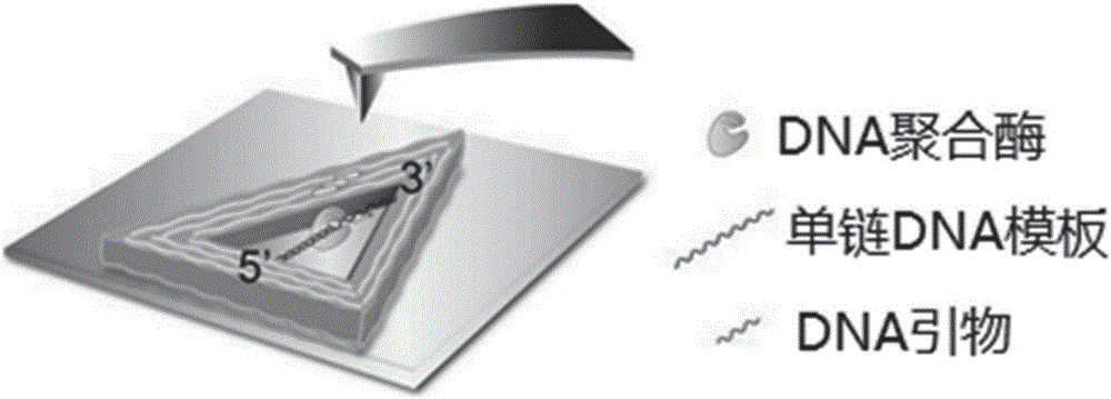
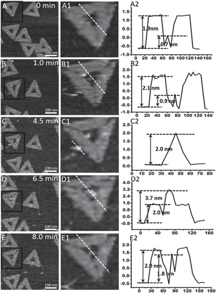
[0044] Add 3.0 μL of the triangular single-stranded DNA-hollow DNA origami prepared in Example 1 dropwise on the surface of the newly dissociated mica and let it stand for 3 minutes. After adding 30 μL of TAE buffer to the liquid pool, use an atomic force microscope to scan and image to obtain clear DNA Image of origami nanostructures and DNA template strands, modeled as figure 2 shown. Subsequently, a mixed solution containing 2 μL of DNA polymerase Klenow fragment at a concentration of 85 U / mL and 2 μL of dNTPs at a concentration of 62.5 μM was added to the liquid pool, and scanned and imaged immediately. Scanning adopts "J" scanning head, "tap" mode, and adjusts scanning parameters to form images in the small force area (F<200pN). Atomic force microscope images of samples were collected at 1 min, 4.5 min, 6.5 min and 8 min after adding the mixture...


PUM

PropertyMeasurementUnit
Elastic coefficientaaaaaaaaaa

Abstract

The invention discloses an image visualization method for detecting single molecule DNA duplication. The method comprises the steps that single-chain DNA and a buffering system of a DNA polymerase Klenow fragment combined with the single-chain DNA are scanned with an atomic force microscope, and DNA duplication is subjected to imaging observation through the atomic force microscope, wherein the single-chain DNA contains dNTP, and the two ends of the single-chain DNA are fixed to two different positions on hollow DNA folded paper respectively. According to the method, the DNA duplication process is tracked on the single molecule level, the whole process of combination of the single-chain DNA and single DNA polymerase, moving between the DNA chain and the DNA polymerase and conversion from the single-chain DNA chain to a double-chain DNA chain is recorded, various forms of state distribution of the DNA chain on the surface of a substrate and the changing situation in the interaction process of DNA and the DNA polymerase can be captured, the biomolecule behavior action mechanism can be understood more easily, and data on the time scale and the space scale in the molecule event obtaining process can be obtained more easily.

Description

technical field [0001] The invention relates to the field of nanotechnology, in particular to an image visualization method for detecting single-molecule DNA replication. Background technique [0002] DNA replication mainly refers to the process of using a DNA single strand as a template strand to synthesize another DNA single strand according to the principle of AT and CG pairing. In the body, DNA replication is very important and is closely related to life processes such as the occurrence, development and evolution of organisms. In vitro, many modern biotechnologies are also involved, including DNA sequencing, forensic examination, nanobiosensor detection, and widely used DNA amplification technology. [0003] DNA replication is a biomolecular reaction process. Using advanced fluorescence resonance energy transfer (FRET) technology and optical tweezers technology, many dynamic process information of DNA replication reactions, such as DNA replication speed, have been obtai...

Claims


Application Information

Patent Timeline
IPC IPC(8): G06F19/26G16B45/00
CPCG16B45/00
Inventor 虞国凯张萍李宾周星飞王奇
Owner SHANGHAI INST OF APPLIED PHYSICS - CHINESE ACAD OF SCI
Why Eureka
Try Eureka