
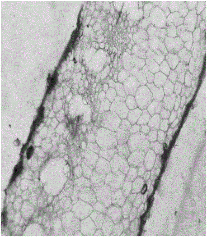
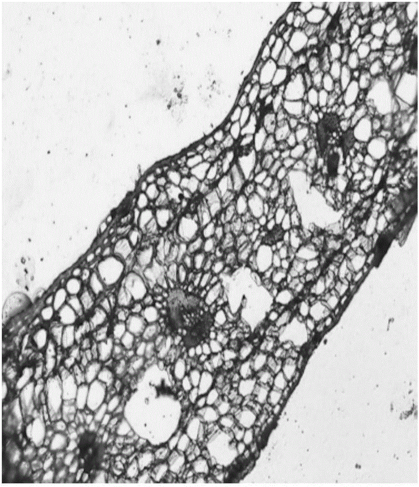
Specimen making method for observing anatomic structure of lamina of tillandsia

An anatomical structure and specimen making technology, applied in the field of leaf specimen making, can solve problems such as difficulty in application, and achieve the effects of improving the softening process, the operation method is simple, and the embedding operation procedure is simplified

Active Publication Date: 2016-10-26
JIANGSU POLYTECHNIC COLLEGE OF AGRI & FORESTRY
View PDF6 Cites 5 Cited by
  • Summary
  • Abstract
  • Description
  • Claims
  • Application Information

AI Technical Summary

Problems solved by technology

[0004] However, the leaf structure of air pineapple is very different from that of conventional plants, and the research methods of conventional leaf anatomy are difficult to apply to it.

Method used


Image

  • Specimen making method for observing anatomic structure of lamina of tillandsia
  • Specimen making method for observing anatomic structure of lamina of tillandsia

Examples


Embodiment 1

[0030] 1. Blade selection

[0031] Select a single plant with no pests and diseases and good growth conditions, and select healthy leaves in the middle of the plant. Choose the breed as Overlord.

[0032] 2. Material fixation:

[0033] Different varieties of leaves have different sizes, depending on the size of the leaves, select a specification, cut the leaves into small rectangular pieces of 3mm×5mm, 4mm×6mm, 5mm×7mm or 6mm×8mm, wash the cut materials with clean water and put them into FAA fixative For medium fixation, the volume of fixative was 20 times that of the fixed tissue. FAA fixative configuration: 70% or 50% ethanol 90mL: glacial acetic acid: formaldehyde = 18:1:1, fixed for 30 hours.

[0034] 3. Material softening:

[0035] 1) Put the sample into a 1:1 solution of 50% glycerin + 50% ethanol (95%) and soak for 25 days.

[0036] 2) Put the material into a mixture of hydrogen peroxide and propionic acid (1:1), take out the sample with tweezers every 30 minutes t...

Embodiment 2

[0060] The selected species is Tillandsia twigs.

[0061] During the material fixing in step 2, the fixing time is 24 hours;

[0062] Step 3: During material softening, put the sample into a 1:1 solution of 50% glycerin+50% ethanol (95%), and soak for 20 days; the volume of hydrogen peroxide and propionic acid in the mixed solution is 2:1;

[0063] Step 6: Infiltrate the material into wax, and place it in an electric thermostat incubator at 37°C for 24 hours after wax sealing;

[0064] In Step 9, put the slide glass in an electric thermostat incubator at 37°C for 2 days.

Embodiment 3

[0066] The selected variety is tricolor Tillandsia.

[0067] During the material fixing in step 2, the fixing time is 36 hours;

[0068] Step 3: During material softening, put the sample into a 1:1 solution of 50% glycerin+50% ethanol (95%), and soak for 30 days; the volume of hydrogen peroxide and propionic acid in the mixed solution is 1:1;

[0069] Step 6: Infiltrate the material into the wax, and place it in an electric thermostat incubator at 37°C for 48 hours after the wax sealing is completed;

[0070] In Step 9, the material is attached to the slide, and the slide is placed in a 37°C electric constant temperature incubator for 5 days.


PUM


Abstract

The invention discloses a specimen making method for observing an anatomic structure of a lamina of tillandsia. The specimen making method for observing the anatomic structure of the lamina of the tillandsia comprises the following steps of (1), selecting the lamina; (2), fixing a material; (3), softening the material; (4), dehydrating and transparentizing the material; (5), sealing the material with wax; (6), infiltrating the material with wax; (7), embedding the material; (8), trimming and slicing the material; (9), sticking the material; (10), dyeing the material; (11), sealing and observing the material. In comparison with the prior art, according to the specimen making method for observing the anatomic structure of the lamina of the tillandsia, a softening process is improved; a new softening reagent combination is used; aiming at the lamina of the tillandsia, an ideal softening effect can be obtained. In addition, an embedding process is innovated; an operation method is simple and convenient; a complete wax stone can be obtained; an original embedding operating procedure is simplified. The problem that a clear observation material of the anatomic structure of the lamina of the tillandsia cannot be obtained in the prior art is solved.

Description

technical field [0001] The invention relates to a specimen preparation method for observing the anatomical structure of the air pineapple leaf, and belongs to the technical field of leaf specimen preparation. Background technique [0002] Air pineapple (Tillandsia), native to the tropical and subtropical regions of Central and South America, belongs to the bromeliaceae (Bromeliaceae) perennial herb, of which the Tillandsia genus is the main one. Due to its unique ornamental and value, my country began to introduce this species in the early 21st century, and the air pineapple gradually emerged in garden art and environmental protection. [0003] The water and nutrients needed for the growth of air pineapple come from the air without the help of soil, and the leaves, as the part most exposed to the air, are bound to become the main organs for absorbing water and nutrients, and the structure is the basis of function. Therefore, there must be a special structure inside the leav...

Claims


Application Information

Patent Timeline
IPC IPC(8): G01N1/28G01N1/36
CPCG01N1/2813G01N1/36
Inventor 方敏彦章明杨鹤同汪善锋席刚俊
Owner JIANGSU POLYTECHNIC COLLEGE OF AGRI & FORESTRY
Learn More