
Eye ground imaging method based on Android

An imaging method and fundus image technology, applied in ophthalmoscopes, eye testing equipment, medical science, etc., to achieve the effects of protecting eyes, reducing injuries, and omitting the steps of smearing

Inactive Publication Date: 2016-11-09
JIANGSU UNIV
View PDF9 Cites 9 Cited by
  • Summary
  • Abstract
  • Description
  • Claims
  • Application Information

AI Technical Summary

Problems solved by technology

At the same time, Android's default low-light photo mode is completely unsuitable for fundus imaging

Method used


Image

  • Eye ground imaging method based on Android
  • Eye ground imaging method based on Android
  • Eye ground imaging method based on Android

Examples


Embodiment Construction

[0026] The present invention will be further described below in conjunction with the accompanying drawings and specific embodiments, but the protection scope of the present invention is not limited thereto.

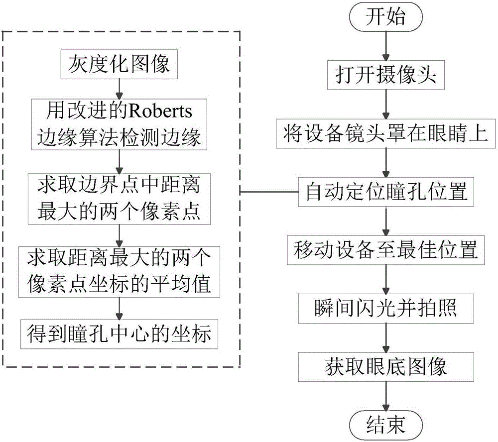
[0027] like figure 1 Shown, a kind of fundus imaging method based on Android, comprises steps:

[0028] In step (1), the hand-held fundus camera is in the photographing interface, and the camera is turned on, and the infrared light is turned on at this time.

[0029] In step (2), the device lens of the hand-held fundus camera is covered on the eye to be detected, and the eye can be previewed by infrared light irradiation.

[0030] Step (3), adjusting the position of the hand-held fundus camera, and automatically detecting the pupil position through an image processing algorithm;

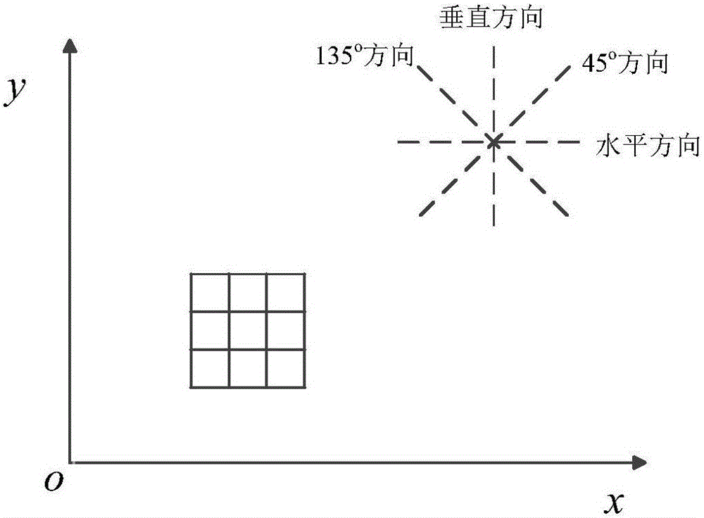
[0031] 1), the camera shoots a fundus image I[x,y], establishes a Cartesian coordinate system xoy with the lower left point of the image as the origin O, and performs grayscale processing on t...


PUM


Abstract

The invention discloses an eye ground imaging method based on Android. The eye ground imaging method includes the steps that when an eye ground image is photographed, an infrared light and an image preprocessing algorithm are used for positioning the pupil to find the optimal position for image photographing; when the photographing key is pressed, instant flashing and photographing are carried out to obtain the high-definition eye ground image. The default logic of photographing under dark light of Android is changed, mydriasis through drug smearing or high light illumination on the eye is not needed during photographing, damage to the eye by a traditional eye ground imaging method is effectively avoided, while operation is convenient, the high-definition image is obtained, and the eye ground condition of a user can be conveniently judged better.

Description

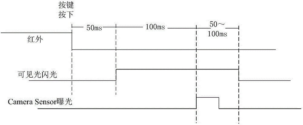
technical field [0001] The invention belongs to the field of fundus imaging, in particular to an Android-based fundus imaging method. Background technique [0002] In low-light or no-light environments, the traditional Android system has a default low-light camera function. First, turn on the camera, click to take a photo, the flash is turned on, and a pre-flash is performed; then, the flash is automatically turned off, and after a short time, the flash and the Camera Sensor (camera sensor) exposure are synchronized to take pictures to obtain the best brightness photos. [0003] The fundus is an important part of the sensory organ of the eye, and most of the lesions of the eyes come from the fundus. At the same time, there are a large number of various arteries and veins in the fundus. Through the observation and analysis of the choroid and retina of the fundus, people can make early diagnosis and prevention of many diseases, such as arteriovenous thrombosis, diabetes, arte...

Claims


Application Information

Patent Timeline
Patent Type & Authority Applications(China)
IPC IPC(8): A61B3/14A61B3/12
CPCA61B3/12A61B3/14
Inventor 许桢英朱建栋洪红张琦许冉冉
Owner JIANGSU UNIV
Learn More