Multi-gamma-photo simultaneous medicine emission time coincidence nuclear medical imaging system and method

A gamma photon and imaging system technology, applied in the field of nuclear medicine imaging, to achieve the effect of reducing the total count, improving the signal-to-noise ratio, and simplifying the image reconstruction algorithm

Active Publication Date: 2016-11-16
TSINGHUA UNIV
View PDF4 Cites 24 Cited by
  • Summary
  • Abstract
  • Description
  • Claims
  • Application Information

AI Technical Summary

Problems solved by technology

[0005] The purpose of the present invention is to solve the above-mentioned PET and SPECT problems in principle. The present...

Method used


Image

  • Multi-gamma-photo simultaneous medicine emission time coincidence nuclear medical imaging system and method
  • Multi-gamma-photo simultaneous medicine emission time coincidence nuclear medical imaging system and method
  • Multi-gamma-photo simultaneous medicine emission time coincidence nuclear medical imaging system and method

Examples


Embodiment Construction

[0020] A multi-gamma photon simultaneous emission drug time conforming nuclear medicine imaging system and method proposed by the present invention is described in detail in conjunction with the accompanying drawings and embodiments as follows:

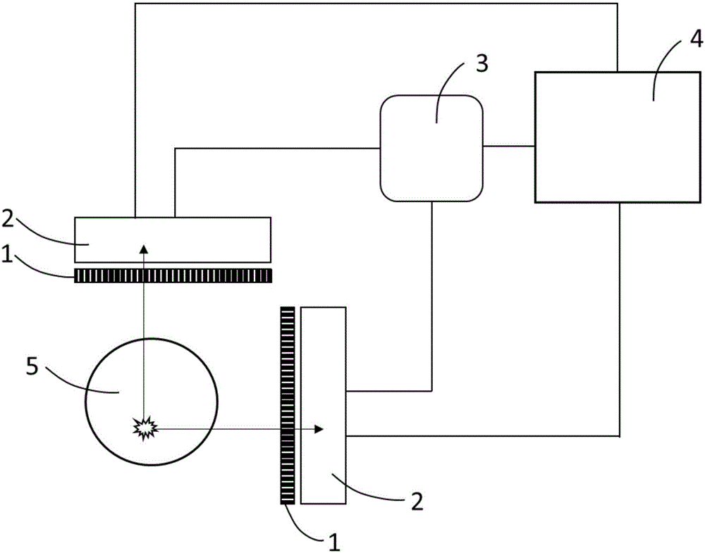
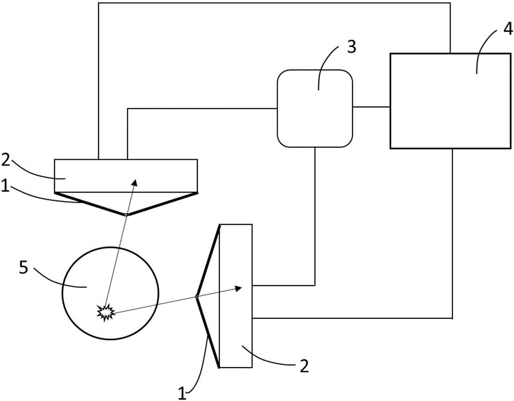
[0021] The overall structure of the imaging system of this embodiment is as follows figure 1 As shown, it consists of two detector probes arranged perpendicular to each other on the detection planes, a time coincidence module 3 and a computer platform 4, and each detector probe consists of a parallel hole collimator 1 and a gamma photon detector with time measurement function 2 configuration; wherein each parallel hole collimator 1 is respectively placed on the front end of the corresponding gamma photon detector 2 so that the multi-gamma photons generated by the decay of the radionuclide in the imaging object 5 can only be along the plane perpendicular to the gamma photon detector Directional emission can be detected by the gamma pho...


PUM


Abstract

The invention discloses a multi-gamma-photo simultaneous medicine emission time coincidence nuclear medical imaging system and method, and belongs to the field of nuclear medical images. The system comprises multiple detector probes arranged in non-parallel mode, a time coincidence module and a computer platform. Each detector probe is composed of a collimator and a gamma photo detector with a time measuring function. Multiple gamma photos radiated by detection radionuclides within a short time form multiple gamma-photo coincidence events. The point with the smallest sum, of distances of projection lines determined by the multiple gamma-photo coincidence events, calculated by the method is the disintegration position of radionuclides, and the distribution of radionuclides in living bodies can be obtained by accumulating a certain number of mult-gamma-photo coincidence events. By means of the imaging system and method, reconstruction algorithm is simplified, the signal to noise ratio of reconstructed images is improved, the requirement for the gamma photo total count is lowered, and the irradiation risk to patients is lowered.

Description

technical field [0001] The invention belongs to the field of nuclear medicine imaging, and in particular relates to a nuclear medicine imaging system and method for simultaneous emission of drugs by multiple gamma photons. Background technique [0002] Nuclear medicine imaging technology is one of the important technical means for disease diagnosis in modern medicine. It uses radionuclide-labeled molecules to participate in the physiological metabolic process of organisms, detects X-rays or gamma photons emitted by radionuclide outside the body and reconstructs the image to obtain the distribution of radionuclide, so that it can be used non-invasively It can be used to observe the organ metabolism of organisms and obtain diagnostic information of diseases. In the field of nuclear medicine imaging, the two most important imaging systems are Positron Emission Tomography (PET) and Single-Photon Emission Computed Tomography (SPECT). Both PET and SPECT have been widely used in ...

Claims


Application Information

Patent Timeline
IPC IPC(8): A61B6/03
CPCA61B6/037A61B6/4266A61B6/5205A61B6/54A61B6/03A61B6/06A61B6/5282G01T1/161G01T1/17G01T1/29
Inventor 刘亚强马天予范鹏
Owner TSINGHUA UNIV
Why Eureka
Try Eureka