Fusion protein disulfide bond pairing analysis method

A technology of fusion protein and analysis method, which is applied in the direction of analysis materials, material separation, instruments, etc., can solve the problems of many types of enzyme digestion, inconvenient, and insufficient fragment ions, and achieve the effect of rich fragment ions and few enzyme digestion methods

Active Publication Date: 2020-11-10
SUNSHINE GUOJIAN PHARMA (SHANGHAI) CO LTD
View PDF4 Cites 0 Cited by
  • Summary
  • Abstract
  • Description
  • Claims
  • Application Information

AI Technical Summary

Problems solved by technology

For the confirmation of disulfide bond pairing in the prior art, multiple enzyme digestion methods can be used in combination with MS / MS mode to confirm 5 pairs of disulfide bond pairing (K J Leicester et al., compositions and methods for producing compositions , Chinese Patent Publication No.: CN101448852), but there are relatively many types of enzyme digestion in this method, and the disadvantages such as fragment ions are not abundant
[0004] rhCTLA4-Ig is a modified Fc (hinge, CH2 and CH3 domains) linking the extracellular domain of human cytotoxic T-lymphocyte-associated antigen 4 (CTLA-4) to human immunoglobulin G1 (IgG1) A fusion protein formed from above, which has been developed by BMS for the treatment of diseases such as rheumatoid arthritis (common name abatacept, trade name ), the current disulfide bond analysis methods for fusion proteins such as rhCTLA4-Ig often need to be confirmed by multiple enzyme digestions, which is very inconvenient

Method used


Image

  • Fusion protein disulfide bond pairing analysis method
  • Fusion protein disulfide bond pairing analysis method
  • Fusion protein disulfide bond pairing analysis method

Examples


Embodiment 1

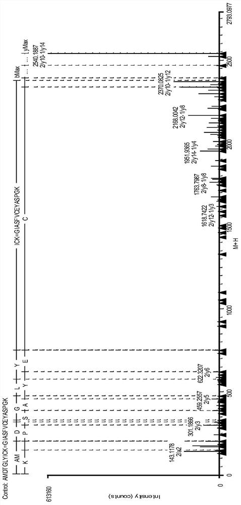
[0045] Example 1: Fusion protein disulfide bond pairing analysis (trypsin digestion)

[0046] Fusion protein disulfide bond pairing analysis steps:

[0047] (1) Removal of N-Glycosylations

[0048] Take 1mg rhCTLA4-Ig fusion protein sample, add 50mM NH 4 HCO 3 (pH 8.0) solution to a volume of 20-50 μL, add 1 μL of peptide N-glycosidase PNGase F (NEB, 500000u / mL), mix well, and incubate at 37°C for 24 hours.

[0049] (2) Protein denaturation treatment

[0050] Take 0.5 mg of the protein sample incubated in (1) and add it to 380 μl of denaturation buffer (8M guanidine hydrochloride+5mM EDTA+0.5M Tris, pH8.3), mix well, and incubate at 37°C for 60 minutes. Ultrafiltration centrifugation or desalting column desalting to 50mM NH 4 HCO 3 (pH8.0) buffer.

[0051] (3) Enzymolysis

[0052] Take 100 μg of the protein in (2), add trypsin to digest the antibody protein at 1:25 (wt / wt), and incubate at 37°C for 4 hours.

[0053] (4) Reduction and termination of enzymatic hydrolysi...

Embodiment 2

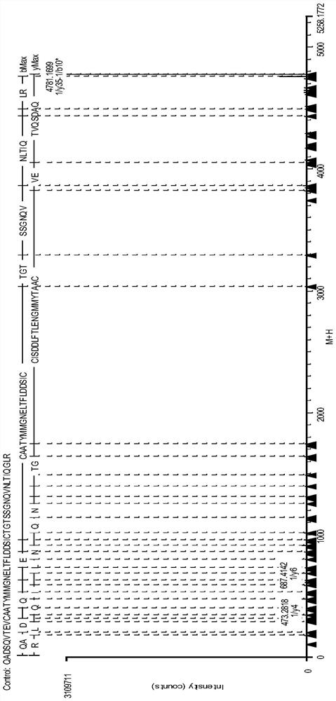
[0068] Example 2: Analysis of the complete molecular weight of the fusion protein (trypsin combined with chymotrypsin digestion)

[0069] Fusion protein disulfide bond pairing analysis steps:

[0070] (1) Removal of N-Glycosylations

[0071] Take 1mg rhCTLA4-Ig fusion protein sample, add 50mM NH 4 HCO 3 (pH8.0) solution to a volume of 20-50 μL, add 1 μL of peptide N-glycosidase PNGase F (NEB, 500000u / mL), mix well, and incubate at 37°C for 24 hours.

[0072] (2) Protein denaturation treatment

[0073] Take 0.5mg of the protein sample incubated in (1) and add it to 380ul denaturation buffer (8M guanidine hydrochloride+5mM EDTA+0.5M Tris, pH8.3), mix well, and incubate at 37°C for 60 minutes. Ultrafiltration centrifugation or desalting column desalting to 50mM NH 4 HCO 3 (pH8.0) buffer.

[0074] (3) Enzymolysis

[0075] Take 100 μg of the protein in (2), add trypsin at 1:25 (wt / wt), add chymotrypsin at 1:50 (wt / wt) to digest the protein, and incubate at 37°C for 4 hours....


PUM


Abstract

The invention relates to an analysis method for disulfide bond pairing of fusion protein. The method comprises the following steps: 1) removing N-sugar of the fusion protein; 2) subjecting the fusion protein without N-sugar to denaturation; 3) subjecting the denatured fusion protein to enzymatic hydrolysis with a specific enzyme under non-reduction conditions; 4) dividing the fusion protein having undergone enzymatic hydrolysis into two samples, subjecting one of the samples to reduction while the other not to reduction and terminating enzymatic hydrolysis of the two samples; and 5) after termination of enzymatic hydrolysis, subjecting disulfide bond peptide fragments of the samples to peptide mass fingerprinting. The method provided by the invention overcomes steric hindrance action of N-sugar during enzymatic hydrolysis of the protein with the specific enzyme and inhibitory effect of N-sugar on ionization efficiency of the peptide fragments, reduces enzyme digestion manners and can accurately confirm and locate the disulfide bond peptide fragments.

Description

technical field [0001] The invention relates to the field of biotechnology, in particular to a protein disulfide bond pairing analysis method. Background technique [0002] The disulfide bond is the S-S bond, which is a covalent bond between sulfur atoms in the form of -S-S- formed by the oxidation of two sulfhydryl groups. Cysteine ​​plays a very important role in forming a stable protein spatial structure, maintaining the correct spatial conformation, and regulating biological activity. The correct pairing of disulfide bonds is conducive to the rapid folding of the peptide chain and the formation of a tight and stable spatial structure, forming a local hydrophobic center, which can prevent water molecules from entering the interior of the peptide to destroy hydrogen bonds and form a stable higher-order structure region. [0003] The disulfide bond is only relatively stable, and its covalent bond is easily reduced and broken. When the disulfide bond is broken and reduced t...

Claims


Application Information

Patent Timeline
Patent Type & Authority Patents(China)
IPC IPC(8): G01N30/02G01N30/06
Inventor 吕锋华环民霞刘周阳谭青乔
Owner SUNSHINE GUOJIAN PHARMA (SHANGHAI) CO LTD
Why Eureka
Try Eureka