
Imaging device and method as well as PET/CT (positron emission tomography/computed tomography) imaging device

An imaging method and imaging device technology, applied in the field of medical diagnosis, capable of solving the problems of small scanning range of CT imaging unit 11 and imaging of patients, etc., achieving the effects of facilitating image fusion, simple registration, and easy realization

Inactive Publication Date: 2016-12-07
SHANGHAI UNITED IMAGING HEALTHCARE
View PDF5 Cites 17 Cited by
  • Summary
  • Abstract
  • Description
  • Claims
  • Application Information

AI Technical Summary

Problems solved by technology

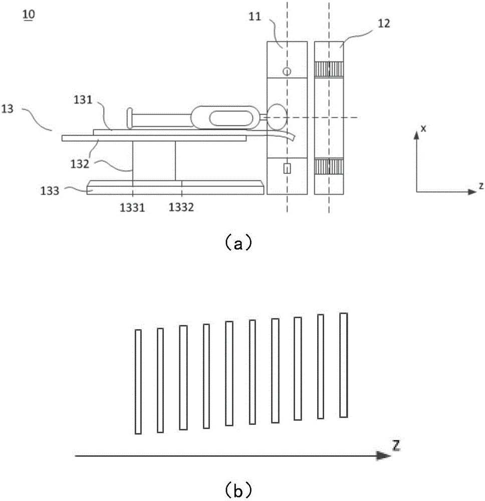
Since the patient cannot be imaged before the bed board 131 moves to the CT scanning surface, the farther the distance between the initial position of the bed board 131 and the CT scanning surface is, the greater the scanning range of the CT imaging unit 11 is when the stroke of the bed board 131 is constant. smaller

Method used


Image

  • Imaging device and method as well as PET/CT (positron emission tomography/computed tomography) imaging device
  • Imaging device and method as well as PET/CT (positron emission tomography/computed tomography) imaging device
  • Imaging device and method as well as PET/CT (positron emission tomography/computed tomography) imaging device

Examples


Embodiment Construction

[0064] In order to make the above objects, features and advantages of the present invention more comprehensible, specific implementations of the present invention will be described in detail below in conjunction with the accompanying drawings.

[0065] Many specific details are set forth in the following description to facilitate a full understanding of the present invention, but the present invention can also be implemented in other ways than described here, so the present invention is not limited by the specific embodiments disclosed below.

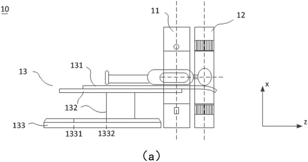
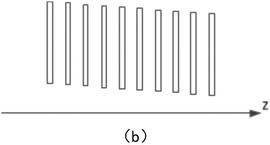
[0066] The present invention provides an imaging device, which determines the linear relationship between the height of the bed board and the moving distance of the bed board in the axial direction according to the previously acquired images, and adjusts the bed board according to the linear relationship so that the surface of the bed board is inclined and conforms to the linear relationship , the imaging scan is carried out under this c...


PUM


Abstract

The invention relates to the field of medical imaging and discloses an imaging method and device. The imaging device comprises an acquiring unit for acquiring images, a determining unit, a height adjusting unit and an imaging unit for executing imaging scanning, wherein the determining unit is used for determining the linear relation between the bed board height in the images and the moving distance of a bed board in the length direction of the bed board; the height adjusting unit is used for adjusting the bed board according to the linear relation, so that the surface of the adjusted bed board meets the linear relation. The imaging method and device can compensate the difference between the bed board sinkage in different imaging modes and facilitate image fusion.

Description

technical field [0001] The invention relates to the field of medical diagnosis, in particular to a PET / CT imaging device. Background technique [0002] With the advancement of science and technology, medical imaging technology has made great progress, and there are more and more imaging modes, such as X-ray imaging (X-Ray Photography), magnetic resonance imaging (Magnetic Resonance Imaging, referred to as MRI), computed tomography ( Computed Tomography (abbreviated as CT), positron emission tomography (abbreviated as PET), etc., different imaging modes have their own strengths and complement each other. [0003] For example, PET scanning is a relatively advanced clinical examination imaging technology in the field of nuclear medicine. It uses positron nuclide as a tracer, and the functional metabolic state of the lesion can be understood through the uptake of the tracer by the lesion site, so as to make a correct diagnosis of the disease. PET can display the pathophysiologi...

Claims


Application Information

Patent Timeline
IPC IPC(8): A61B6/03A61B6/04
CPCA61B6/03A61B6/037A61B6/0407A61B6/0487
Inventor 刘伟平刘士涛
Owner SHANGHAI UNITED IMAGING HEALTHCARE
Learn More