Multiple organ segmentation method based on deep convolutional neural network and regional competition model

A convolutional neural network and neural network technology, applied in the direction of instruments, image analysis, image data processing, etc., can solve the problem of not getting accurate segmentation results

Active Publication Date: 2016-12-07
ZHEJIANG DE IMAGE SOLUTIONS CO LTD
View PDF6 Cites 49 Cited by
  • Summary
  • Abstract
  • Description
  • Claims
  • Application Information

AI Technical Summary

Problems solved by technology

The latest algorithm uses a convolutional neural network to detect multi

Method used


Image

  • Multiple organ segmentation method based on deep convolutional neural network and regional competition model
  • Multiple organ segmentation method based on deep convolutional neural network and regional competition model
  • Multiple organ segmentation method based on deep convolutional neural network and regional competition model

Examples


Embodiment Construction

[0062] The present invention will be further described in detail below in conjunction with the drawings and specific embodiments:

[0063] The following embodiments can enable the professionals in the field to understand the present invention more comprehensively, but do not limit the present invention in any way.

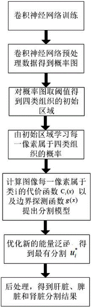
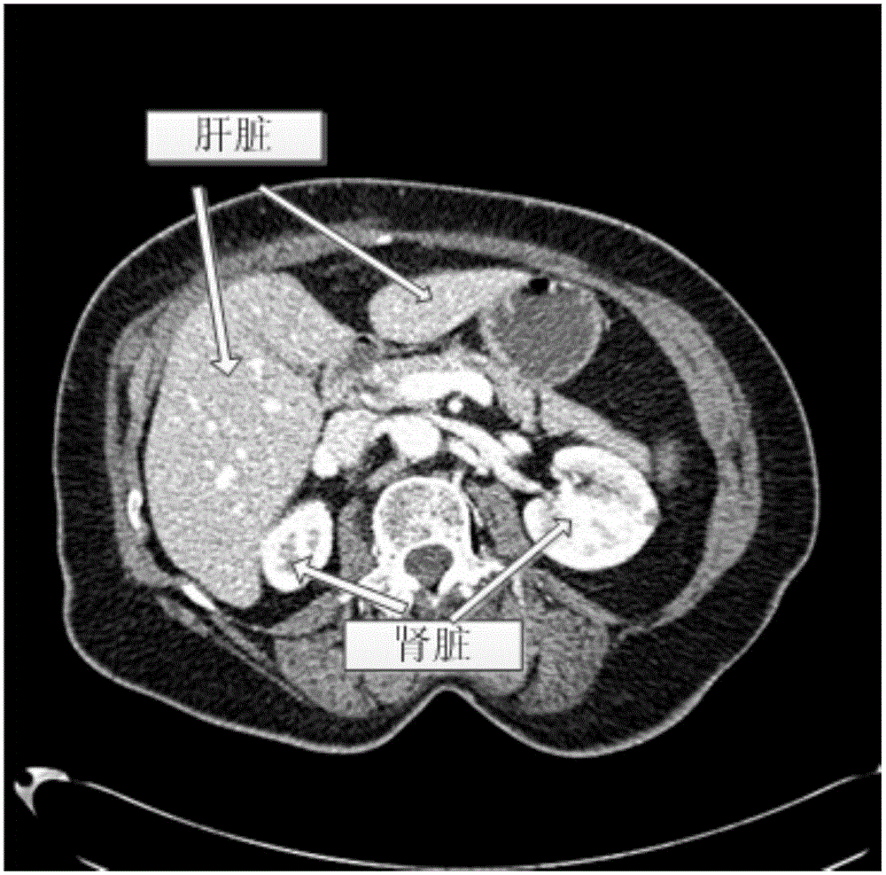
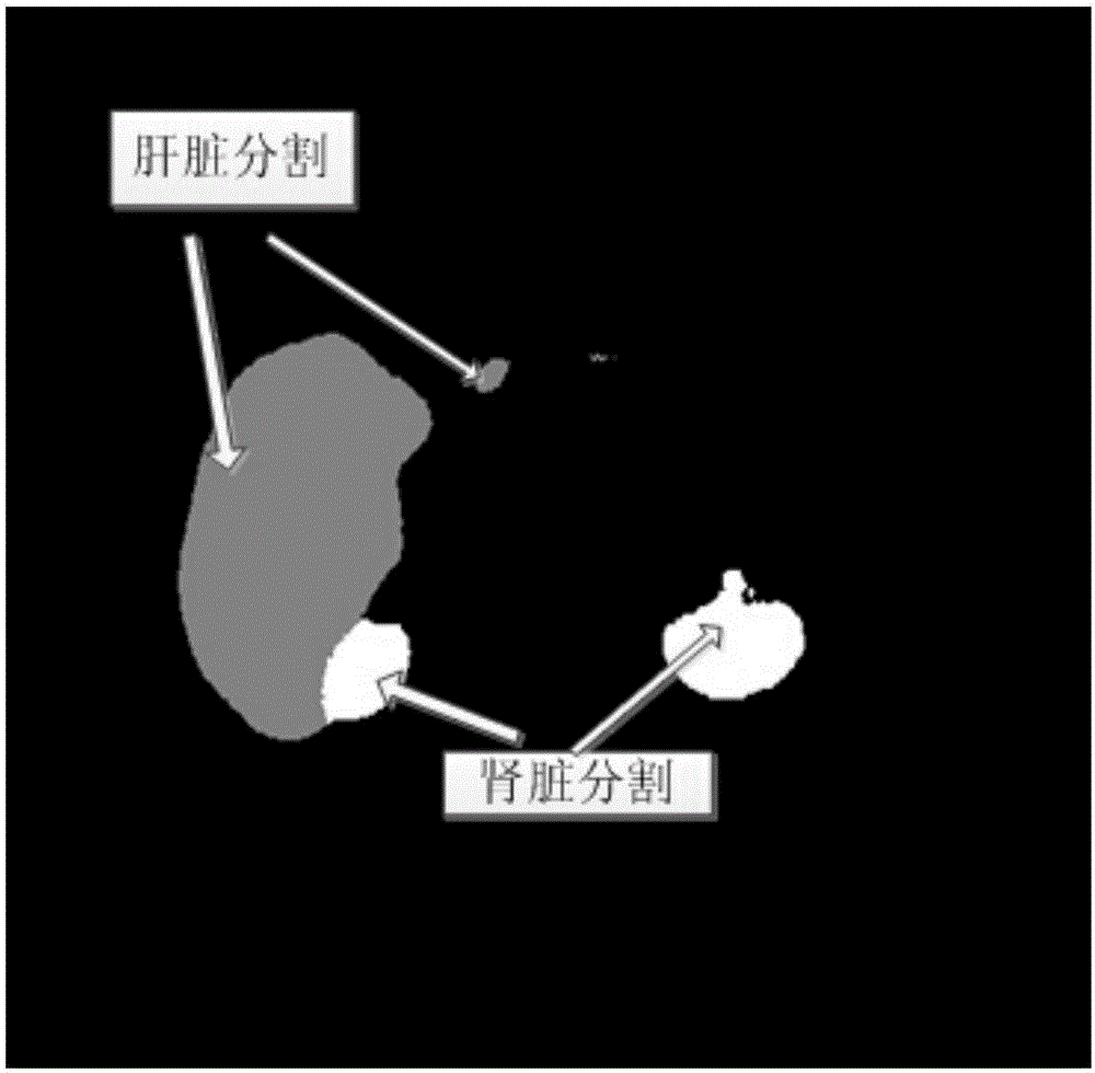
[0064] Such as figure 1 As shown, the multi-organ segmentation method based on convolutional neural network and regional competition model is used to simultaneously segment the liver, spleen and kidney in the computed tomography angiography image. The specific steps are as follows:

[0065] The process one specifically includes the following steps:

[0066] Step A: Collect 140 abdominal liver CTA volume data with a size of 512×512×N, and the doctor will give the liver segmentation standard results of these data, where N is the number of layers of volume data. For data with N>286, delete the number of layers without liver tissue in the data, so that the number of data laye...


PUM


Abstract

The invention relates to medical image processing and aims to provide a multiple organ segmentation method based on a deep convolutional neural network and a regional competition model. The multiple organ segmentation method based on the deep convolutional neural network and the regional competition model comprises processes of training a three-dimensional convolutional neural network; using the trained three-dimensional convolutional neural network to learn prior probability images of liver, spleen, kidney and background in CTA volume data; determining an initial segmentation region of each tissue according to the prior probability image of each tissue; determining the probability of each pixel point belonging to each of the four tissues in the image; establishing a multiple region segmentation model based on regional competition; solving the model using the convex optimization method; and performing post-processing to obtain the contour of each organ. The invention uses the convolutional neural network to automatically and rapidly detect positions of liver, spleen and kidney at the belly, thereby obtaining the prior probability image of each organ is obtained. Then the invention uses the regional competition model, so that the contours of liver, spleen and kidney can be accurately segmented.

Description

Technical field [0001] The invention relates to the field of medical image processing, in particular to a multi-organ segmentation method based on a deep convolutional neural network and a regional competition model. Background technique [0002] The segmentation of abdominal organs has important research significance and clinical value. In clinical practice, doctors often use CT machines, that is, computed tomography scanners, to obtain a series of flat gray-scale tomographic images of the human abdomen, and to judge the size, position, and relationship of various organs by continuously viewing these images. Fast and automatic segmentation of organs from CT images is the first step in visualization. More important than visualization is that determining the location and area of ​​the organ of interest is of great significance in radiotherapy surgery. Only according to the spatial geometry and volume of the organs can an accurate radiotherapy plan and radiotherapy dose be formul...

Claims


Application Information

Patent Timeline
IPC IPC(8): G06T7/00
CPCG06T2207/10081G06T2207/20081G06T2207/20084G06T2207/30056
Inventor 孔德兴胡佩君吴法
Owner ZHEJIANG DE IMAGE SOLUTIONS CO LTD
Why Eureka
Try Eureka