
Method for extracting lung lobe contour from DR image

A contour extraction and lung lobe technology, applied in the field of medical informatization, can solve problems such as delayed treatment, physical harm to patients, missed diagnosis of lung cancer, etc., and achieve the effect of reducing usage rate, improving accuracy, and reducing visual load

Active Publication Date: 2017-02-15
SICHUAN UNIV
View PDF5 Cites 13 Cited by
  • Summary
  • Abstract
  • Description
  • Claims
  • Application Information

AI Technical Summary

Problems solved by technology

This is because some lung cancers and tuberculosis are difficult to distinguish on imaging, and it is very easy to misdiagnose, mistreat or delay treatment clinically.
There are some clinical patients who obviously only have pulmonary tuberculosis, but are diagnosed with lung cancer by a single primary hospital based on an imaging examination, and even blindly started radiotherapy and chemotherapy, which caused serious harm to the patient's body.
On the contrary, there are also some patients who obviously have lung cancer combined with pulmonary tuberculosis, but are misdiagnosed as lung cancer, and are only treated as simple pulmonary tuberculosis.

Method used


Image

  • Method for extracting lung lobe contour from DR image
  • Method for extracting lung lobe contour from DR image
  • Method for extracting lung lobe contour from DR image

Examples


Embodiment Construction

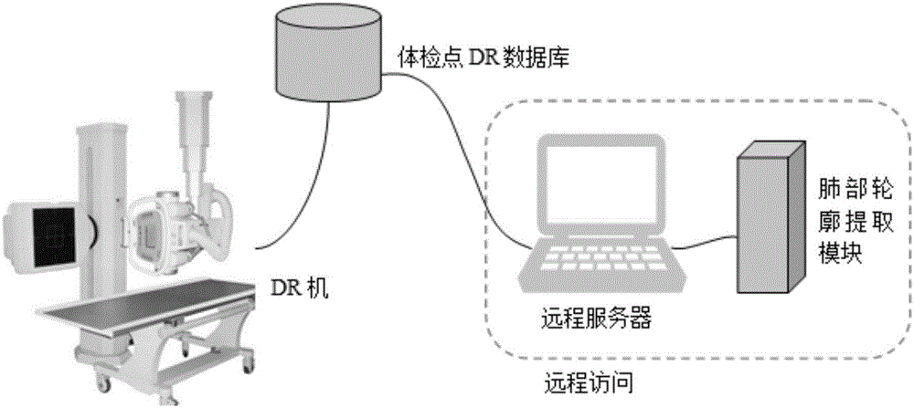
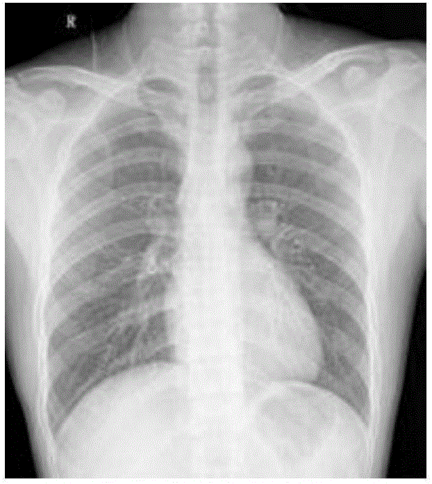
[0038]The present invention will be further described in detail below in conjunction with the accompanying drawings and specific embodiments. The functions of the present invention include: reading digital chest X-ray image DICOM data (Digital Imaging and Communications in Medicine, medical digital imaging and communication), utilizing digital image processing technology to detect the contour of the lungs, and determining its area range; The image of the lung area that distinguishes the shadow of the lung lesion, the processing result is confirmed by the doctor to ensure reliability, and the feedback information from the doctor can improve the system performance online. The basic idea of ​​the method of the present invention is as follows: the method for automatically suppressing the lungs and ribs facing the DR film of the chest X-ray is applied to the basic large-scale physical examination point and the screening of major infectious diseases, and is mainly composed of compute...


PUM


Abstract

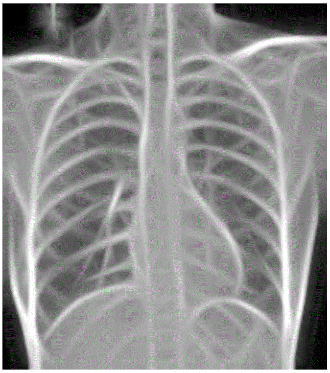
The invention discloses a method for extracting a lung lobe contour from a DR image. The method comprises the following steps: a representative template of a lung lobe contour is obtained through offline training; a chest DR image lung lobe area extraction system is initialized; according to the size of a DICOM image, the image is subjected to three-layer pyramid decomposition; a Gabor filter set is used to reconstruct the to-be-processed image, and the residual error of the reconstructed image after Gabor filter is converted into a black and white image; the black and white image is refined with a Zhan-Suen refinement algorithm; with each offline training template called as a convolution kernel operator, the contour image is subjected to convolution; a local optimal convolution value of the optimal possibility is filtered out of the convolution results and subjected to combined evaluation; and a lung lobe contour shape is generated by combining the most matching upper and lower templates and the most matching positions. The method improves the work efficiency and inspection precision of lung disease inspection by doctors, supports further deepening the informatization of tuberculosis monitoring, and facilitates popularization of regular resident infectious disease examination screening of tuberculosis.

Description

technical field [0001] The invention relates to the field of medical informatization, in particular to a method for extracting lung lobe contours in diagnosis assistance for large-scale screening of tuberculosis infectious diseases. Background technique [0002] DR film is a digital photographic image, which is the abbreviation of Digital Radiography. Chest DR film images are a substitute for traditional chest fluoroscopy imaging, which has high imaging resolution and low radiation. X-ray imaging is the main method of physical examination to screen for lung diseases, including lung inflammation, mass, tuberculosis, lung cancer, etc. Chest x-ray DR film uses different densities of human tissues to image differences under X-rays to observe lesions in areas with small differences in thickness and density. However, the structure of human tissue is complex. The thorax and abdominal cavity contain key organs of the human body, including various organs of high and low density. T...

Claims


Application Information

Patent Timeline
Patent Type & Authority Applications(China)
IPC IPC(8): G06K9/62G06K9/46
CPCG06V10/443G06V2201/07G06F18/23213A61B6/5217G06T2207/20016G06T2207/20081G06T7/74A61B6/50G06T7/0014G06T2207/10116G06T2207/30061G16H50/30G06T7/13A61B5/7225A61B6/5211
Inventor 王俊峰唐鹏姬郁林梁宗安
Owner SICHUAN UNIV
Learn More