Dry eye disease full-automatic detecting apparatus

A fully automatic and detector technology, which is applied in the fields of eye testing equipment, medical science, diagnosis, etc. It can solve the problems of easy loss of the Placido cone, long duration of the test process, troublesome operators, etc., and achieves reliable light source and brightness. The effect of adjusting, reducing the difficulty of inspection, and shortening the inspection time

Active Publication Date: 2017-03-22
SUZHOU INST OF BIOMEDICAL ENG & TECH CHINESE ACADEMY OF SCI
View PDF11 Cites 5 Cited by
  • Summary
  • Abstract
  • Description
  • Claims
  • Application Information

AI Technical Summary

Problems solved by technology

The traditional tear film break-up time (BUT) test is the time when the first dry spot appears on the cornea under a slit lamp microscope after corneal fluorescein sodium staining. The accuracy of the test results depends on the experience of the observer and the effect of the stain on the tear film. Stability; tear secretion test uses standard test paper for tear measurement, gently put two standard test papers into the junction of the middle and outer 1 / 3 of the conjunctival sac under the test eye, the patient looks down or gently closes the eyes, and removes the test paper after 5 minutes , read the scale of the test paper after leaving it for one minute, and judge the diagnosis result according to the test value. This method can also test the basic value under the state of surface anesthesia of the patient, so as to avoid the tear secretion caused by the stimulation of the test paper itself from affecting the accuracy of the test result. The test method is also one of the commonly used methods in clinical ophthalmology at present. The cost is low, but the test process lasts for a long time, and the invasive method brings some discomfort to the patient; Keratograph5M can comprehensively analyze dry eye symptoms, but the equipment is relatively large , poor portability, high cost, poor popularity
[0004] At the same time, the black stripe part of the Placido ring in the imaging component of the existing dry eye analyzer is printed with ordinary ink. After the tear film breakup time is measured, the Placido cone needs to be removed, and then the tear river height measurement, lipid layer image For acquisition and meibomian gland image acquisition, the Placido cone needs to be installed again after the three measurements are completed. This design mode will undoubtedly bring some trouble to the operator, and the Placido cone is easy to lose after it is removed.

Method used


Image

  • Dry eye disease full-automatic detecting apparatus
  • Dry eye disease full-automatic detecting apparatus
  • Dry eye disease full-automatic detecting apparatus

Examples


Embodiment 1

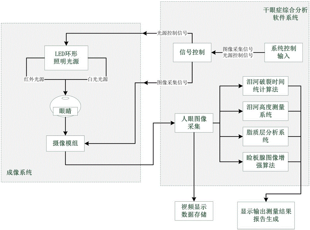
[0039] Such as figure 1 What is shown is an implementation form according to the present invention, which includes: a probe assembly for taking pictures of the patient's eyes, a handle integrated with the probe assembly, a portable device for carrying the probe assembly, and performing multiple symptom analysis on the images The analysis system and the terminal for outputting images, data and reports.

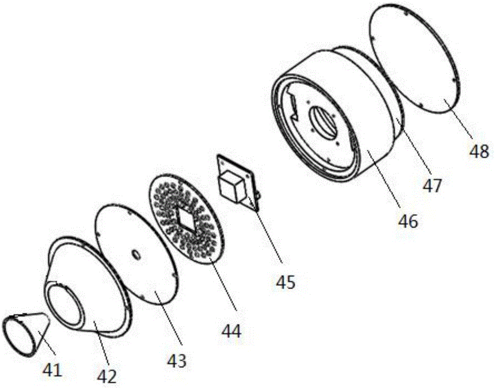
[0040] In the present invention, the probe assembly 4 is an integrated structure consisting of an imaging assembly, an illumination source 44, a camera module 45, a camera fixing device 46, a control board 47 and a cover plate 48, such as figure 2 As shown, the imaging assembly, the lighting source 44 and the camera module 45 are fixed on one side of the camera fixing device 46, and the control board 47 is fixed on the other side of the camera fixing device 46. A complete structure, the imaging assembly includes a tapered tube 41, a fixed sleeve 42 and a filter 43 arranged in...

Embodiment 2

[0053] In this embodiment, the handle is a hollow structure, the connecting wire of the probe assembly 4 passes through the handle and is connected to the analysis device, and the handle can be separated from the portable device to become an independent handheld analysis device; The headrest assembly is arranged behind the probe assembly, and the adjustable workbench assembly and the front, rear, left, and right distances of the probe assembly relative to the headrest assembly can be adjusted by swinging the adjustable hand lever front, rear, left, and right, and the headrest can be adjusted by rotating the adjustable hand lever up and down. The up and down position of the assembly's chin rest relative to the probe assembly. Specifically, the lower end of the handle is set on the adjustable table assembly 3 through a detachable part, and the headrest assembly 1 is arranged behind the probe assembly 4. When the patient is positioned on the headrest assembly 1, the hand can be ad...

Embodiment 3

[0056] On the basis of Embodiment 1, when the probe assembly is removed to become an independent handheld device, for the convenience of operation, the first tear film break-up time check button, the first tear river height measurement button, the second A meibomian gland image acquisition and enhancement button, the first lipid layer analysis button, each button corresponds to the corresponding measurement mode, each measurement mode corresponds to an illumination mode and imaging mode, when one of the buttons is selected, the illumination source The lighting mode of 44 is automatically switched to the corresponding lighting mode, and at the same time, the camera module 45 automatically adjusts the imaging mode and performs autofocus to perform high-definition imaging of the patient's eyes. Specifically, the PC and the handle are respectively provided with buttons for controlling the measurement mode, each button corresponds to a corresponding measurement mode, each measuremen...


PUM


Abstract

The invention discloses a dry eye disease full-automatic detecting apparatus, which comprises a probe assembly, a handle, a portable device and an analysis device, wherein the probe assembly comprises an imaging assembly, a lighting source, a camera module, a camera fixing device, a control board and a cover plate; the imaging assembly comprises an optical filter, a fixed sleeve and a conical drum; the conical drum is of an annular structure chequered with black and white; black rings are formed by conducting silk-screen printing with oil ink, and the oil ink is transmissive in infrared light and is non-transmissive in white light; one end of the handle is connected to the lower end of the probe assembly; the portable device comprises a base, a head supporting assembly, an adjusting handle rod and an adjustable workbench assembly; and the analysis device comprises an image data acquisition unit, a data detection unit and a data analysis unit. According to the detecting apparatus provided by the invention, the black annular structures on the conical drum is invisible to some infrared light while is visible to the white light, so that the demands of four imaging modes are satisfied; and it is unnecessary to detach the Placido conical drum when the detecting apparatus is used, so that a using process is simplified and the conical drum is prevented from getting lost easily.

Description

technical field [0001] The invention relates to a dry eye syndrome detection device, in particular to a dry eye syndrome automatic detection instrument. Background technique [0002] Dry eye refers to the general term for various diseases characterized by abnormal tear quality and quantity or dynamic abnormalities caused by any reason, accompanied by ocular discomfort, resulting in various diseases characterized by ocular surface tissue lesions. Conjunctival xerosis. Common symptoms include dry eyes, easy fatigue, itchy eyes, foreign body sensation, burning sensation, sticky secretions, fear of wind, photophobia, and sensitivity to external stimuli. A survey in the United States shows that 10%-15% of adults suffer from dry eye, 17% in Japan, 10.3% in Australia, and the prevalence of dry eye in my country is about 21%-33.7%. The incidence of dry eye is increasing year by year. With the widespread use of electronic screens, environmental degradation, abuse of eye drops, weari...

Claims


Application Information

Patent Timeline
Patent Type & Authority Applications(China)
IPC IPC(8): A61B3/14A61B3/10
CPCA61B3/0008A61B3/101A61B3/14
Inventor 董月芳付威威周哲陈奭
Owner SUZHOU INST OF BIOMEDICAL ENG & TECH CHINESE ACADEMY OF SCI
Why Eureka
Try Eureka