Quantum dot targeting probe kit for detecting tumor of colon cancer
A technology of quantum dots and kits, applied in the field of quantum dot targeted probe kits, can solve problems such as poor photostability, easy photobleaching, and inability to display lesions.
- Summary
- Abstract
- Description
- Claims
- Application Information
AI Technical Summary
Problems solved by technology
Method used
Image

Examples
Embodiment 1
[0020] Embodiment 1 Composition and configuration of a quantum dot targeting probe kit for detecting colon cancer tumors:
[0021] The kit includes a) TCP-1-H6 polypeptide, b) TP-1-H6 polypeptide, c) water-soluble quantum dot, d) FITC, e) DAPI.
[0022] A). The polypeptide sequence of TCP-1-H6 is cyclo(1,9)-CTPSPFSHCGP 9 G 2 h 6 .
[0023] B). The sequence of TP-1-H6 polypeptide is TPSPFSHGP 9 G 2 h 6 . Both peptides were synthesized by solid phase synthesis.
[0024] C). The water-soluble quantum dots are quantum dots containing Zn, such as CdSe / ZnS, CdTe / ZnS, CdSe / ZnSe or CdTe / ZnSe. In the quantum dots, glutathione is used as a modifier to convert fat-soluble quantum dots into water-soluble quantum dots.
[0025] D). Fluorescein isothiocyanate (FITC)
[0026] E).4,6-diamidino-2-phenylindole (DAPI)
Embodiment 2
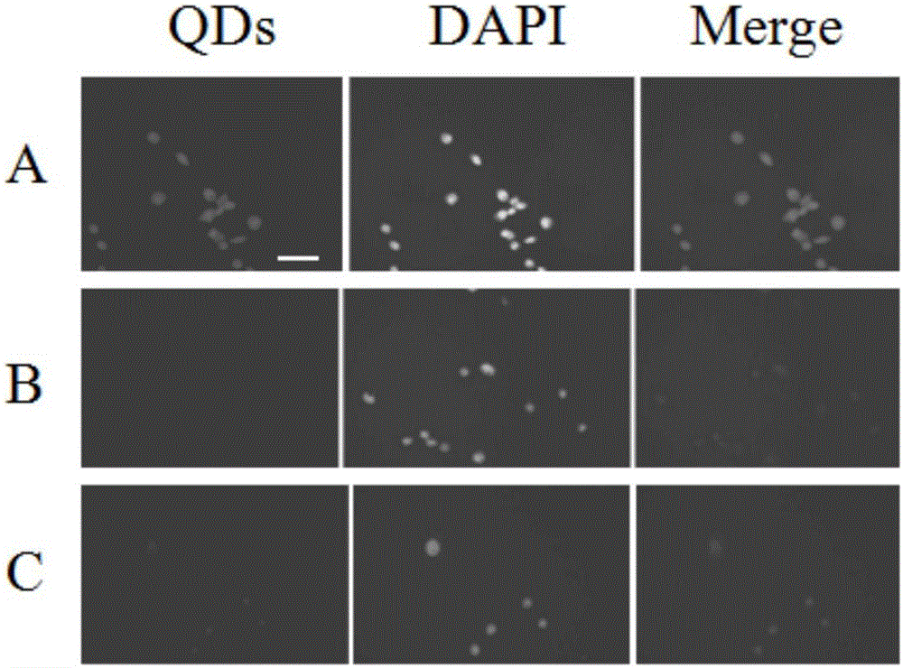
[0027] Embodiment 2 detects Colon26 cells with a quantum dot targeting probe kit for detecting colon cancer tumors, and the process is as follows:
[0028] 1. Mix the peptide and quantum dots at a ratio of 18:1, shake for 10 minutes, and remove excess peptide molecules by ultrafiltration to obtain a quantum dot-polypeptide fluorescent probe.
[0029] 2. Colon 26 cells whose nuclei were stained with DAPI were cultured in RPMI-1640 (Invitrogen) medium and incubated overnight in a carbon dioxide incubator (5% CO2, 37°C).
[0030] 3. After TCP-1-H6-QD was mixed, it was incubated at 37°C for 4 hours, and the unlabeled peptide quantum dots and TP-1-H6-QD were used as a control.
[0031] 4. The incubated cells were washed three times with PBS buffer (pH 7.4).
[0032] 5. Observe the fluorescence imaging under a Nikon C1si laser confocal fluorescence microscope (Nikon, Japan) (excitation wavelength is 488nm).
[0033] The experimental results confirmed that the fluorescence of quant...
Embodiment 3
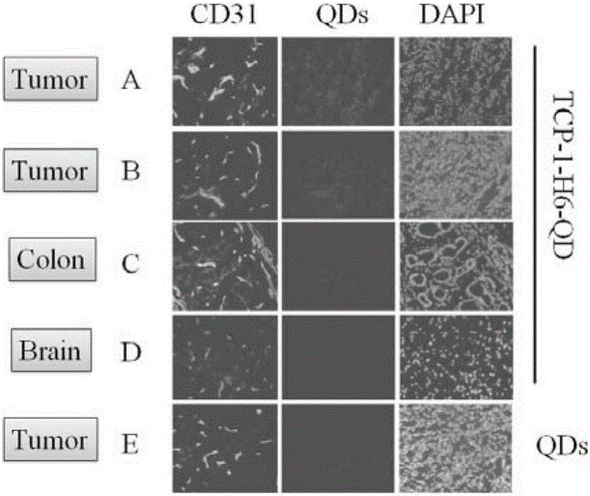
[0034] Example 3 Using a quantum dot targeting probe kit for detecting colon cancer tumors to detect mouse colon cancer tumor tissue sections, the process is as follows:
[0035] 1. Mix the polypeptide and quantum dots at a ratio of 18:1, shake for 10 minutes, and remove excess polypeptide molecules by ultrafiltration to obtain the quantum dot-polypeptide fluorescent probe.
[0036] 2. Select colon cancer tumor tissue slices from mice containing colon cancer tumors, and select normal colon tissue and brain tissue as controls.
[0037] 3. The blood vessels in the selected tissue were stained with FITC-labeled secondary antibody, and the nuclei were stained with DAPI.
[0038] 4. After mixing tumor tissue or other tissues with TCP-1-H6-QD (100nM), incubate at 37°C for 1h.
[0039] 5. The incubated tissue was washed three times with PBS buffer (pH 7.4).
[0040] 6. Observe the fluorescence imaging under a Nikon C1si laser confocal fluorescence microscope (Nikon, Japan) (excitat...
PUM
 Abstract
Description
Claims
Application Information
 - R&D
- Intellectual Property
- Life Sciences
- Materials
- Tech Scout
- Unparalleled Data Quality
- Higher Quality Content
- 60% Fewer Hallucinations
Browse by: Latest US Patents, China's latest patents, Technical Efficacy Thesaurus, Application Domain, Technology Topic, Popular Technical Reports.
© 2025 PatSnap. All rights reserved.Legal|Privacy policy|Modern Slavery Act Transparency Statement|Sitemap|About US| Contact US: help@patsnap.com