

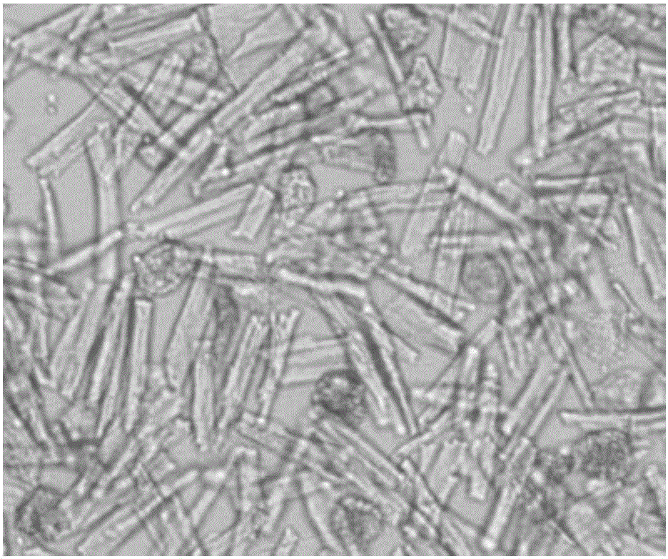
Myocardial cell separation method
A separation method and cardiomyocyte technology, applied in cell dissociation methods, animal cells, vertebrate cells, etc., can solve the problems of reducing the survival rate of cardiomyocytes, deformation of cardiomyocytes, etc., and achieve the effect of avoiding changes and improving activity
- Summary
- Abstract
- Description
- Claims
- Application Information
AI Technical Summary
Problems solved by technology
Method used
Image

Examples
Embodiment 1
[0041] A method for separating cardiomyocytes of adult rats, comprising the following steps:
[0042] Step 1: Put the freshly isolated heart into the pre-cooled protective solution at 4°C, cut out the ventricular muscle tissue, wash it with washing solution, and then cut the ventricular muscle tissue into small pieces of 1mm×1mm×1mm with ophthalmic scissors , then soaked in the flushing solution for 3 minutes, filtered with gauze to obtain a myocardial tissue sample, and set aside;
[0043] Each liter of the protective solution contains the following components: 8g of NaCl, 0.4g of KCl, 0.06g of Na 2 HPO 4 ·H2 O, 0.06 g of KH 2 PO 4 , 0.35g of NaHCO 3 , the solvent is double distilled water;
[0044] Each liter of washing solution contains the following components: 9g of NaCl, 0.3g of KCl, and the solvent is double distilled water.
[0045] Step 2, the myocardial tissue sample was placed in 1 g / L trypsin solution, digested at 37° C. for 8 min, and the supernatant was dis...
Embodiment 2
[0051] A method for separating cardiomyocytes from neonatal rats, comprising the following steps:
[0052] Step 1: Put the freshly isolated heart into the pre-cooled protective solution at 4°C, cut out the ventricular muscle tissue, wash it with washing solution, and then cut the ventricular muscle tissue into small pieces of 1mm×1mm×1mm with ophthalmic scissors , then soaked in the flushing solution for 5 minutes, filtered with gauze to obtain a myocardial tissue sample, and set aside;
[0053] Each liter of the protective solution contains the following components: 8g of NaCl, 0.4g of KCl, 0.06g of Na 2 HPO 4 ·H 2 O, 0.06 g of KH 2 PO 4 , 0.35g of NaHCO 3 , the solvent is double distilled water;
[0054] Each liter of washing solution contains the following components: 9g of NaCl, 0.3g of KCl, and the solvent is double distilled water.
[0055] Step 2, the myocardial tissue sample was placed in 1 g / L trypsin solution, digested at 37° C. for 5 min, and the supernatant ...
PUM
 Abstract
Description
Claims
Application Information