Lung pneumothorax CT image classified diagnosis method based on machine learning

A technology of CT imaging and machine learning, applied in image analysis, computer components, instruments, etc., to reduce the burden on doctors and improve the accuracy and misdiagnosis rate

Inactive Publication Date: 2017-07-07
杭州健培科技有限公司
View PDF2 Cites 13 Cited by
  • Summary
  • Abstract
  • Description
  • Claims
  • Application Information

AI Technical Summary

Problems solved by technology

[0005] Aiming at the deficiency that the existing CT images of pneumothorax completely rely on the subjective diagnosis of clinicians, the present invention discloses a method for classification and diagnosis of CT images of lung pneumothorax based on machine learning

Method used


Image

  • Lung pneumothorax CT image classified diagnosis method based on machine learning
  • Lung pneumothorax CT image classified diagnosis method based on machine learning
  • Lung pneumothorax CT image classified diagnosis method based on machine learning

Examples


Embodiment Construction

[0036] The preferred embodiments of the present invention will be described below in conjunction with the accompanying drawings. It should be understood that the preferred embodiments described here are only used to illustrate and explain the present invention, and are not intended to limit the present invention.

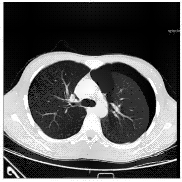
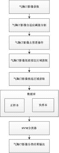
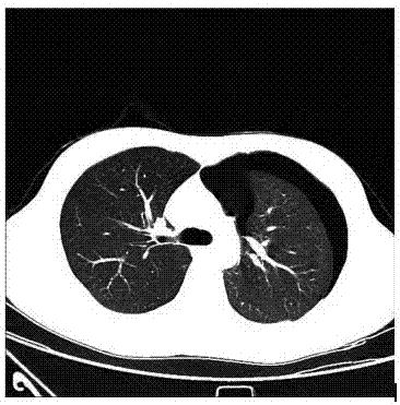
[0037] Such as figure 1 As shown, the method for classification and diagnosis of lung pneumothorax CT images based on machine learning, the steps include: Step 1, obtain pneumothorax CT image data from clinical hospitals and perform pneumothorax area calibration operations, and the calibration area includes the boundary and central point of the pneumothorax area, etc. ; Step 2, perform image processing on the calibrated pneumothorax CT image; Step 3, perform positive and negative sample calibration on the CT image data after image processing, to obtain positive samples and negative samples; Step 4, use the obtained sample data to perform SVM Training prediction diag...


PUM


Abstract

The invention discloses a lung pneumothorax CT image classified diagnosis method based on machine learning. The method comprises the steps that 1, pneumothorax CT image data is obtained from a clinical hospital, and pneumothorax region calibration operation is performed; 2, image processing is performed on the pneumothorax CT image obtained after calibration; 3, positive and negative sample calibration is performed on CT image data obtained after image processing to obtain positive samples and negative samples; 4, obtained sample data is utilized to train an SVM to predict classified diagnosis; and 5, a trained SVM model is utilized to perform classified diagnosis on the lung pneumothorax CT image. By utilizing a machine learning method, clinical doctors are freed out of heavy x-ray plate reading tasks, the burden on the doctors is relieved, and meanwhile the accuracy of diagnosis is greatly improved.

Description

technical field [0001] The invention belongs to the field of medical image big data classification research, in particular to a method for classification and diagnosis of lung pneumothorax CT images based on machine learning. Background technique [0002] With the development of computer technology and medical imaging technology, especially the recent rapid development of machine learning, deep learning and big data technology, it has provided huge technical support for the development of modern medical imaging diagnosis. Therefore, it is necessary to apply these technologies to the computer-aided diagnosis system, and use the powerful advantages of machine learning, deep learning and big data technology to further improve the accuracy and misdiagnosis rate of automatic medical imaging diagnosis, and reduce the burden on clinicians. [0003] Pneumothorax is generally considered to be a state of gas entering the pleural cavity to form a gas accumulation state. There are many ...

Claims


Application Information

Patent Timeline
Patent Type & Authority Applications(China)
IPC IPC(8): G06F19/00G06K9/62G06T7/11G06T7/136
CPCG16H50/20G06T2207/30061G06T2207/10081G06F18/2411G06F18/214
Inventor 苏宝星程国华谢玮宜许卫东季红丽
Owner 杭州健培科技有限公司
Why Eureka
Try Eureka