ELM-based fundus image retinal vessel segmentation method

A technology for retinal blood vessels and fundus images, applied in the field of image processing, can solve problems such as long training time and segmentation time, low accuracy, uneven background fundus images, etc.

Inactive Publication Date: 2017-07-07
CENT SOUTH UNIV
View PDF1 Cites 20 Cited by
  • Summary
  • Abstract
  • Description
  • Claims
  • Application Information

AI Technical Summary

Problems solved by technology

The learning-based retinal vessel segmentation method is the method with the highest accuracy among all methods, but the existing methods are not effective for fundus images with very uneven background, especially fundus images with lesions, and the accuracy is not high. In addition, the training time and the segmentation time is too long, it is difficult to use in practical applications

Method used


Image

  • ELM-based fundus image retinal vessel segmentation method
  • ELM-based fundus image retinal vessel segmentation method
  • ELM-based fundus image retinal vessel segmentation method

Examples


Embodiment 1

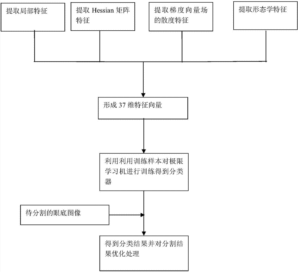
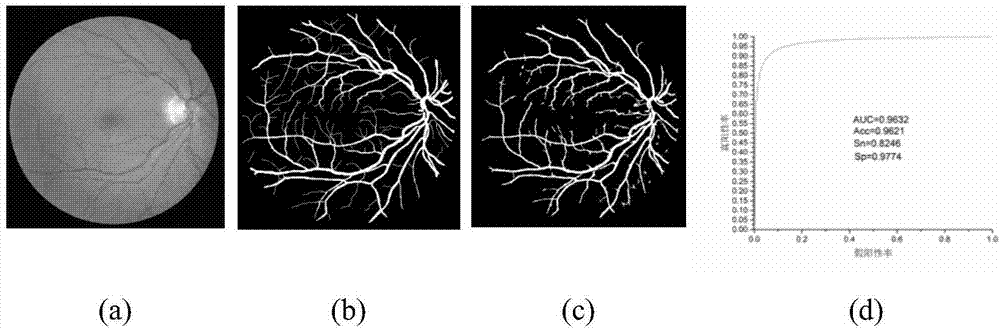
[0109] According to the method described in this article, the figure 2 Figure a is segmented, and the resulting manual marking and segmentation results are shown in Figure b and Figure c, respectively, and the obtained ROC curve is shown in Figure d; from figure 2 We can see the segmentation results, and the ROC curve of the method in this paper (the area between the curve and the X coordinate axis can evaluate the pros and cons of the segmentation algorithm, the larger the area, the better), from the area between the curve and the x axis AZ=0.9632 , it can be seen that the segmentation method in this paper is accurate and credible, and the accuracy reaches 0.9621, the sensitivity reaches 0.8246 and the specificity reaches 0.9774, which better proves that the segmentation method in this paper is accurate and credible.

Embodiment 2

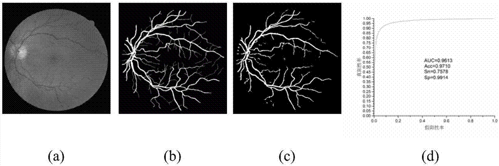
[0111] According to the method described in this article, the image 3 Figure a is segmented, and the resulting manual marking and segmentation results are shown in Figure b and Figure c, respectively, and the obtained ROC curve is shown in Figure d; from image 3 We can see the segmentation results, and the ROC curve of the method in this paper (the area between the curve and the X-axis can evaluate the quality of the segmentation algorithm, the larger the area, the better), from the area between the curve and the x-axis AUC=0.9613 , it can be seen that the segmentation method in this paper is accurate and credible, and the accuracy reaches 0.9710, the sensitivity reaches 0.7578 and the specificity reaches 0.9914, which better proves that the segmentation method in this paper is accurate and credible.

Embodiment 3

[0113] According to the method described in this article, the Figure 4 Figure a is segmented, and the resulting manual marking and segmentation results are shown in Figure b and Figure c, respectively, and the obtained ROC curve is shown in Figure d; from Figure 4 We can see the segmentation results, and the ROC curve of the method in this paper (the area between the curve and the X-axis can evaluate the quality of the segmentation algorithm, the larger the area, the better), from the area between the curve and the x-axis AUC= 0.9602, it can be seen that the segmentation method in this paper is accurate and credible, and the accuracy reaches 0.9673, the sensitivity reaches 0.7601 and the specificity reaches 0.9851, which better proves that the segmentation method in this paper is accurate and credible.

[0114] Depend on Figure 2-Figure 4 The data shows that the accuracy is above 0.9500, the specificity is above 0.9800, and the sensitivity is above 0.7500. All indicators a...


PUM


Abstract

The present invention discloses an ELM-based fundus image retinal vessel segmentation method. According to the method, a 39-dimensional feature vector including a Hessian matrix feature, a local feature, a gradient field feature and a morphological feature is constructed for each pixel in a fundus image so as to be used for judging whether each pixel belongs to pixels on a blood vessel; and training samples are adopted to train an ELM, so that a classifier can be obtained, classification judgment of each pixel on the image to be tested is completed, and final segmentation results are obtained. The method has the advantages of short training time, higher segmentation speed of the fundus image to be tested and better extraction effects of the trunk parts of blood vessels, and is advantageous in the processing of high-brightness lesion areas, is suitable for post-processing, provides intuitive results for the lesions of main blood vessels, is suitable for computer-aided quantitative analysis and disease diagnosis of fundus images and has significant clinical significance for the auxiliary diagnosis of related diseases.

Description

technical field [0001] The invention belongs to the field of image processing, in particular to an ELM-based retinal blood vessel segmentation method for fundus images. Background technique [0002] The color fundus image is the only image that can directly capture the microvascular network of the human body in a non-invasive way. Doctors can clearly observe the optic disc, macula and retinal microvascular network in the fundus through the fundus image. The analysis of blood vessels in fundus images, whether there are changes in shape, diameter, scale, branch angle, and whether there is hyperplasia and exudation, is an important method for diagnosing eye diseases and systemic cardiovascular and cerebrovascular diseases such as diabetes and hypertension. One of the basis. With the rapid increase of fundus image data, if doctors only rely on manual observation and empirical diagnosis, it is not only inefficient but also highly subjective. Therefore, using a computer to autom...

Claims


Application Information

Patent Timeline
Patent Type & Authority Applications(China)
IPC IPC(8): G06T7/155G06T7/136G06K9/62G06N3/04
CPCG06N3/04G06F18/241
Inventor 邹北骥崔锦恺朱承璋张子谦陈瑶
Owner CENT SOUTH UNIV
Why Eureka
Try Eureka