One-dimensional non-focused and focused ultrasonic double array scanning imaging device and method

A non-focused ultrasound and ultrasound array technology, applied in medical science, diagnosis, diagnostic recording/measurement, etc., can solve the problems of inaccurate photoacoustic 3D image reconstruction, lack of two-dimensional array, increased system complexity and cost, etc. , to achieve the effect of convenient operation, high imaging quality and good adaptability

Active Publication Date: 2017-09-01
PEKING UNIV
View PDF7 Cites 7 Cited by
  • Summary
  • Abstract
  • Description
  • Claims
  • Application Information

AI Technical Summary

Problems solved by technology

Since the two-dimensional area array needs to contain many ultrasonic transducer elements and amplification and acquisition circuits, which greatly increases the complexity and cost of the system, there is a lack of large-area two-dimensional area arrays for photoacoustic imaging.
The focused one-dimensional line array can only receive photoacoustic signals in biological tissues within a limited angle, which is not accurate enough for the reconstruction of photoacoustic three-dimensional images.

Method used


Image

  • One-dimensional non-focused and focused ultrasonic double array scanning imaging device and method
  • One-dimensional non-focused and focused ultrasonic double array scanning imaging device and method
  • One-dimensional non-focused and focused ultrasonic double array scanning imaging device and method

Examples


Embodiment 1

[0038] In this embodiment, the tissue to be tested is a small animal with a small size and is suitable for being placed in a water tank.

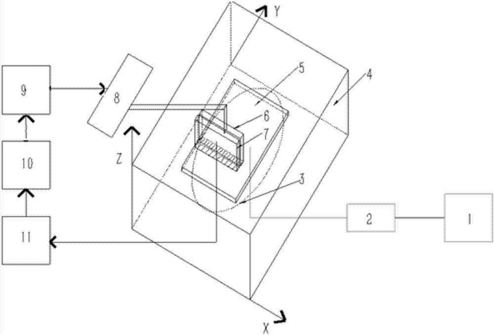
[0039] Such as figure 1 As shown, the dual-array scanning imaging device based on one-dimensional non-focused and focused ultrasound in this embodiment includes: a pulsed laser 1, a refraction divergent element 2, a water tank 4, a carrier plate 5, a non-focused ultrasonic transducer line array 6, an ultrasonic Array 7, electronically controlled translation stage 8, controller 9, computer 10 and data acquisition card 11; wherein, the water tank 4 is filled with water; the non-focused ultrasonic transducer line array 6 and the ultrasonic array 7 are placed parallel to each other back and forth, two The position of the tester is relatively fixed, and it is set in the water tank 4; the tissue to be measured is placed on the object loading plate 5, and the object loading plate 5 is set in the water tank 4; the non-focused ultrasonic transducer ...

Embodiment 2

[0051] Such as Figure 4 As shown, in this embodiment, the tissue to be tested is a relevant part of the human body, such as a breast, and the tissue to be tested is placed on the loading plate 5, and the loading plate 5 is placed under the water tank 4, and the loading plate 5 moves along the z-axis , the sink 4 is pressed on the tissue to be tested. The bottom of the water tank 4 is made of low-density polyethylene, so that an ultrasonic window is formed on the bottom of the water tank, and the ultrasonic window is pressed on the tissue to be tested. Others are the same as embodiment one.


PUM


Abstract

The invention discloses a one-dimensional non-focused and focused ultrasonic double array scanning imaging device and method. The relative positions of a non-focused ultrasonic transducer linear array and an ultrasonic array are fixed and arranged front to back side by side; laser stimulates to-be-tested tissue to generate a photoacoustic signal, the non-focused ultrasonic transducer linear array collects the photoacoustic signal and ultrasonic imaging is carried out on the ultrasonic array at an interval of a laser pulse; the one-dimensional non-focused ultrasonic transducer linear array is scanned and is equivalent to a two-dimensional photoacoustic detection area array, so that the one-dimensional non-focused ultrasonic transducer linear array has a wider receiving angle than a focused linear array; meanwhile, the design and processing difficulty is greatly reduced in comparison with a two-dimensional surface, and the one-dimensional non-focused ultrasonic transducer linear array has great value in animal and photoacoustic tomographic imaging; the image contrast sources of two modes are different and information is complementary, a photoacoustic image and an ultrasonic image are registered and rebuilt to obtain a double-mode three-dimensional imaging result, and can be applied to small animal imaging or tumor disease examination and post-treatment evaluation of a breast cancer and the like.

Description

technical field [0001] The invention relates to an imaging method applied to animals and clinics, in particular to a dual-array scanning imaging device based on one-dimensional non-focused and focused ultrasound and an imaging method thereof. Background technique [0002] Photoacoustic tomography (Photoacoustic tomography, PAT, sometimes referred to as photoacoustic imaging) technology is a method of imaging optical absorbers in tissues by using the effect of ultrasound emitted by tissues after absorbing pulsed laser light. Photoacoustic tomography is a hybrid imaging modality that combines rich optical contrast and high-resolution ultrasound, breaking through the barrier of strong light scattering caused by purely high-resolution optical imaging of tissue, while being different from pure ultrasound imaging , the tissue's ability to absorb photons reflects the molecular composition of the tissue and its function. The fusion of photoacoustic tomography and ultrasound imaging...

Claims


Application Information

Patent Timeline
Patent Type & Authority Applications(China)
IPC IPC(8): A61B5/00
CPCA61B5/0095
Inventor 李长辉张广杰任秋实
Owner PEKING UNIV
Why Eureka
Try Eureka