Multi-arm type intraoperative radiotherapy device

A radiotherapy and robotic arm technology, applied in the field of radiotherapy medicine, can solve problems such as the inability to determine the position, angle, direction, time-consuming, and heavy workload of lesions, and achieve the effect of reducing the burden on clinicians, reducing risks, and saving treatment time

Active Publication Date: 2019-09-03
CANCER INST & HOSPITAL CHINESE ACADEMY OF MEDICAL SCI
View PDF5 Cites 0 Cited by
  • Summary
  • Abstract
  • Description
  • Claims
  • Application Information

AI Technical Summary

Problems solved by technology

[0007] Nevertheless, the above-mentioned technologies still have the following defects. First, there is a lack of three-dimensional stereotaxic technology and devices in clinical applications, and the position of the tumor bed is determined only by visual observation by clinical surgeons and radiologists, or by CT or MRI images before surgery. Unable to accurately determine the position, angle, direction of the lesion, and its relationship with the surrounding normal tissues and vital organs under the condition of the tumor bed exposure, and thus unable to set the optimal path and direction of the electron beam incident, as well as the emission power and irradiation time of the electron beam Wait
Secondly, during clinical treatment, radiotherapy doctors first need to manually place the light-limiting tube on the part of the patient to be irradiated, and then fix it on the operating bed through a fixing device, and then observe the laser positioning mark while moving the radiation head of the accelerator remotely. , so that the central axis of the beam is aligned with the central axis of the light-limiting tube, and finally the treatment is performed. The above steps are complicated and cumbersome, with a large workload and a long time-consuming, which increases the risk of intraoperative radiotherapy, and the positioning accuracy of the radiation head is difficult to guarantee, which is not conducive to Realizing Precise Intraoperative Radiation Therapy

Method used


Image

  • Multi-arm type intraoperative radiotherapy device
  • Multi-arm type intraoperative radiotherapy device
  • Multi-arm type intraoperative radiotherapy device

Examples


Embodiment Construction

[0034] In order to make the technical means, creative features, goals and effects achieved by the present invention easy to understand, the present invention will be further described below in conjunction with specific illustrations.

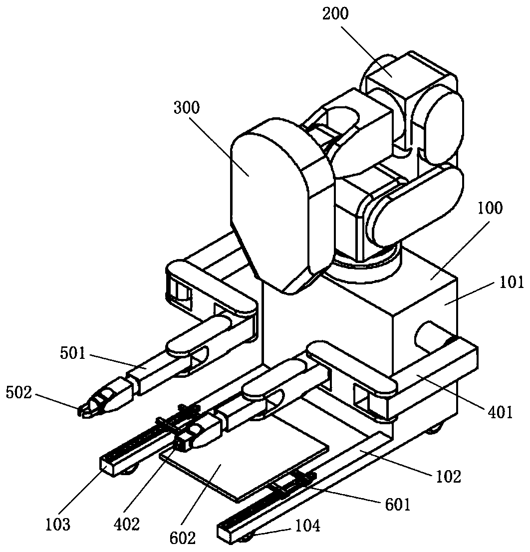
[0035] figure 1 It shows a multi-manipulator intraoperative radiation therapy device in an initial stowed state according to an embodiment of the present invention, so as to facilitate the delivery of the multi-manipulator intraoperative radiation therapy device.

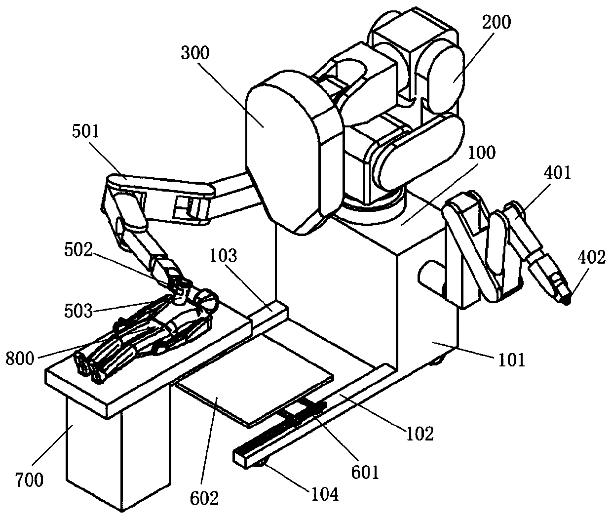
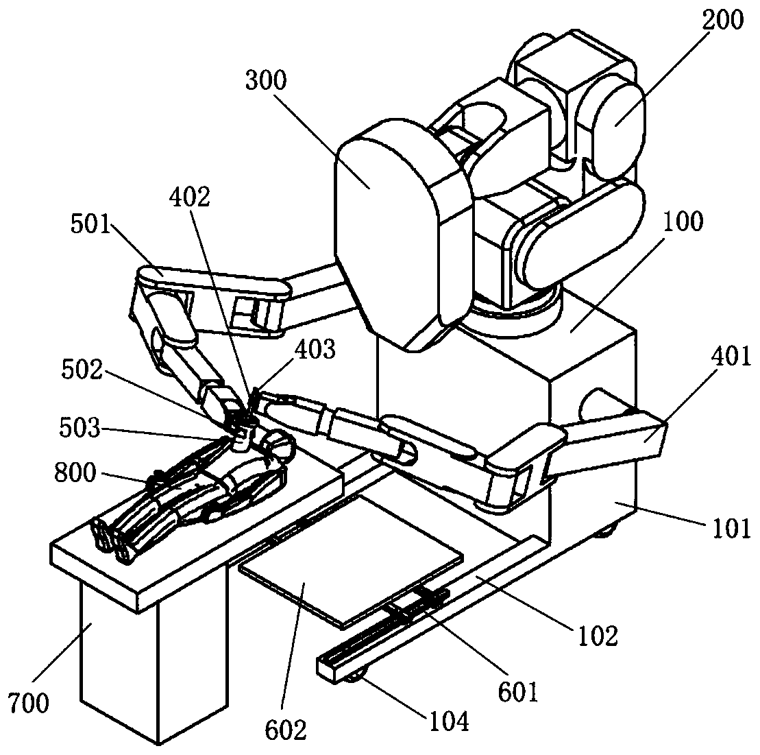
[0036] Such as figure 1 As shown, the multi-manipulator intraoperative radiotherapy device according to an embodiment of the present invention includes: a base 100, a main manipulator 200, a linear accelerator radiation head 300, a left manipulator 401, a left manipulator end gripper 402, The right mechanical arm 501 , the gripper 502 at the end of the right mechanical arm, the two-dimensional motion platform 601 , and the ray blocker 602 .

[0037] The base 100 includes a base body...


PUM


Abstract

The invention relates to a multi-manipulator type intraoperative radiotherapy device, which may include: a base; a main manipulator installed on the base for moving a radiation head installed at its end; a first manipulator installed on the base On the base, there is a first mechanical arm end gripper for clamping imaging equipment or therapeutic light-limiting cylinder; The end gripper of the second robotic arm. The invention can use the robotic arm to drive the imaging device to perform three-dimensional imaging on the tumor bed before intraoperative radiotherapy, and based on this, accurately determine the position of the target area and the relationship between the target area and surrounding normal tissues and important organs, and more accurately determine the irradiation dose and time, which is beneficial to reduce the complications of intraoperative radiotherapy and the toxic side effects caused by radiation. Through the cooperative work of multiple robotic arms and the use of automatic control technology, the workload of clinicians can be greatly reduced, the positioning accuracy can be improved, and it is conducive to the realization of precise intraoperative radiotherapy.

Description

technical field [0001] The present invention generally relates to the field of radiotherapy medicine, and more particularly relates to a multi-manipulator type intraoperative radiotherapy device. Background technique [0002] Intraoperative radiotherapy (intraoperative radiotherapy for short) is one of tumor radiotherapy techniques, which refers to surgical exposure of unresectable lesions after surgical resection of tumor lesions, and treatment of postoperative tumor beds, residual lesions, lymphatic drainage areas or original tumors. For the lesion, a large dose of high-energy electron beam radiation is applied through the surgical field of view. This treatment method can be used to replace traditional external radiation therapy, and can be widely used in the combined treatment of head and neck, chest, abdomen, and pelvic tumors. Early intraoperative radiotherapy was treated with conventional accelerators, which required patients who had been surgically exposed to be move...

Claims


Application Information

Patent Timeline
Patent Type & Authority Patents(China)
IPC IPC(8): A61N5/10
CPCA61N5/1015A61N5/1049A61N5/1083A61N2005/1058A61N5/103A61N2005/1089
Inventor 戴建荣牛传猛马攀李明辉
Owner CANCER INST & HOSPITAL CHINESE ACADEMY OF MEDICAL SCI
Why Eureka
Try Eureka