Method for performing dentition segmentation on cone beam CT image

A CT image and dentition technology, applied in the field of computer vision and image processing, can solve problems such as increasing the burden of data processing

Active Publication Date: 2017-09-26
PEKING UNIV
View PDF5 Cites 19 Cited by
  • Summary
  • Abstract
  • Description
  • Claims
  • Application Information

AI Technical Summary

Problems solved by technology

Atlases and statistical shape models generally come from a large number of CT images,

Method used


Image

  • Method for performing dentition segmentation on cone beam CT image
  • Method for performing dentition segmentation on cone beam CT image
  • Method for performing dentition segmentation on cone beam CT image

Examples


Embodiment Construction

[0075] Below in conjunction with accompanying drawing, further describe the present invention through embodiment, but do not limit the scope of the present invention in any way.

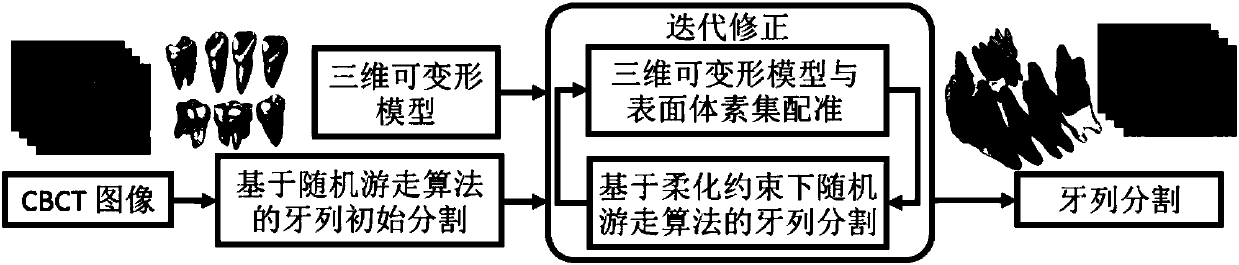
[0076] A CBCT image is a three-dimensional volume image, which is composed of a series of two-dimensional images. In this description, the two-dimensional images are called slice images (layered images). The embodiments of the present invention aim at medical clinical CBCT images, and segment the dentition in the CBCT images based on the graph structure defined in the image area of ​​the body of interest in the CBCT images and a small number of layered images (slice images) interactively marked by users. Among them, a three-dimensional deformable model is used to define the softening constraints, and the dentition segmentation in the volume image is updated through a random walk algorithm based on the softening constraints, and then a reliable segmentation result is obtained by an iterative correction...


PUM


Abstract

The invention discloses a method for performing dentition segmentation on a cone beam CT image (CBCT). The method for performing dentition segmentation on a cone beam CT image comprises steps of defining an image structure according to an interest body image area in the cone beam CT image, obtaining a complete dentition through segmenting the CBCT image on the basis of a semi-supervised random walk algorithm and a soft constraint which is defined and registered by a three-dimensional deformable model, using the three-dimensional deformable model to introduce a soft constraint of a body image for processing noise in body image segmentation based on semi-supervised mark diffusion, adopting an iteration correction method to perform iteration solving problems on mark diffusion under the soft constrain and fitting on a surface voxel set by the three-dimensional model. The method for performing dentition segmentation on the cone beam CT image can effectively eliminate a segmentation error, improves dentition segmentation obtained through single time mark diffusion, improves segmentation accuracy and meets an accuracy requirement for clinic stomatology.

Description

technical field [0001] The invention relates to the technical fields of computer vision and image processing, in particular to a method for segmenting dentition on a cone beam CT (CBCT) image. Background technique [0002] Cone beam computed tomography (CBCT) images are commonly used in maxillofacial and orthodontic surgery for auxiliary surgery prediction and dentition alignment planning. CBCT images can provide patient-specific anatomical information, including complete dentition information. The traditional optical-based methods, such as 3D laser scanning and stereo vision equipment, can only obtain the geometric information of the crown, and the geometric shape of the root cannot be obtained because it is buried in the gum and jawbone. In clinical oral surgery, CBCT images have great advantages over traditional CT imaging techniques due to their low radiation dose and low acquisition cost. However, lower radiation doses and signal-to-noise ratios generally result in po...

Claims


Application Information

Patent Timeline
IPC IPC(8): G06T7/11
CPCG06T2207/10081G06T2207/20104G06T2207/30036
Inventor 裴玉茹艾兴胜查红彬许天民
Owner PEKING UNIV
Why Eureka
Try Eureka