Preparation method of co-crosslinked double network hydrogel scaffold for promoting osteogenic growth
A co-crosslinking and double-network technology, applied in tissue regeneration, medical science, prosthesis, etc., can solve the problems of hydrogel strength decrease, drug release, etc., to promote bone density increase, increase bone formation, and enhance adsorption The effect of the aggregation function
- Summary
- Abstract
- Description
- Claims
- Application Information
AI Technical Summary
Problems solved by technology
Method used
Image
Examples
Embodiment 1
[0044] Example 1 Preparation of Co-crosslinked Double Network Hydrogel Scaffold for Promoting Osteogenic Growth
[0045] The specific steps are:
[0046] 1) Preparation of methacrylic anhydride modified gelatin
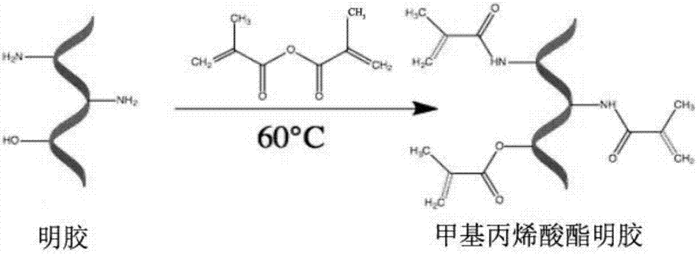
[0047] figure 2 This is the flow chart for the preparation of methacrylic anhydride-modified gelatin. Add 200 mL of phosphate buffer to 20 g of gelatin, and keep stirring at 60°C for 2 hours; Add 1 mL of methacrylic anhydride to the gelatin mixture every 4 minutes while stirring, add 16 times in total, and keep stirring for 2 hours to form methacrylic anhydride-modified gelatin; Place the gelatin solution in 800mL preheated phosphate buffer solution for dilution, and continue to stir slowly for 15 minutes, place the diluted solution in a dialysis bag, the molecular weight cut-off of the dialysis bag is 10KD, soak in pure aqueous solution, Change the dialysate twice a day to remove unreacted methacrylic anhydride, and continue dialysis for one week; after dialysis,...
Embodiment 2
[0054] Example 2 Adhesion and Aggregation Observation of GelMA and GelMA-OGP
[0055] Experimental groups: control group, GelMA group, GelMA-OGP scaffold group.
[0056] Put the GelMA and GelMA-OGP scaffolds into 24-well plates first, then recover MC3T3-E1, centrifuge, count, and plant them on the scaffolds in two 24-well plates (2×10 per well) 4 Cells) (MC3T3-E1 in the control group were directly planted on the culture plate), three replicate wells in each group were added to the prepared α-MEM medium, and cultured in the incubator for 24 hours until the cells fully adhered to the wall. The α-MEM medium was changed every two days, and the control group only changed the α-MEM medium on the culture plate, and cultured for 1 day and 3 days respectively, and observed with live-death staining fluorescence microscope.
[0057] The procedure for live-dead staining is as follows: suck out the culture medium → wash twice with PBS → add 100 ul of the mixture to each well (1 μl of A +...
Embodiment 3
[0059] Example 3 Morphological observation of MC3T3-E1 implanted on the scaffold
[0060] Experimental groups: control group, GelMA group, GelMA-OGP scaffold group.
[0061] Put the GelMA and GelMA-OGP scaffolds into 96-well plates first, then resuscitate, centrifuge, count and plant MC3T3-E1 on the scaffolds in 96-well plates (2×10 per well 3 Cells) (control group MC3T3-E1 were directly planted on the culture plate), added α-MEM medium, and cultured in the incubator for 24 hours, until the cells fully adhered to the wall and grew. The α-MEM medium was replaced every other day (the control group only replaced the α-MEM medium on the culture plate). On the 3rd day, Dapi and phalloidin staining were performed for fluorescence microscope observation.
[0062] Dapi and phalloidin staining steps: Aspirate the culture medium → wash with PBS → fix with paraformaldehyde for 15 minutes → wash with PBS for 2 minutes*5 times → 1ul phalloidin + 300ul PBS (1:300) dilution → 96-well plat...
PUM
| Property | Measurement | Unit |
|---|---|---|
| Concentration | aaaaa | aaaaa |
| Mwco | aaaaa | aaaaa |
Abstract
Description
Claims
Application Information
 - R&D
- Intellectual Property
- Life Sciences
- Materials
- Tech Scout
- Unparalleled Data Quality
- Higher Quality Content
- 60% Fewer Hallucinations
Browse by: Latest US Patents, China's latest patents, Technical Efficacy Thesaurus, Application Domain, Technology Topic, Popular Technical Reports.
© 2025 PatSnap. All rights reserved.Legal|Privacy policy|Modern Slavery Act Transparency Statement|Sitemap|About US| Contact US: help@patsnap.com


