Semi-automatic medical image segmentation method based on shape constraint of point distance function

A distance function and medical image technology, applied in the field of medical image processing, can solve problems such as difficult access to database, single shape, time-consuming manual hand drawing, etc.

Active Publication Date: 2017-10-10
NANJING FORESTRY UNIV
View PDF8 Cites 3 Cited by
  • Summary
  • Abstract
  • Description
  • Claims
  • Application Information

AI Technical Summary

Problems solved by technology

[0016] The above literature has more or less published some similar methods, but the existing prior shapes are either obtained through the analysis of the collected target object shape, or a large number of shape parameters need to be optimized during the solution process; however The collected target shapes are manually drawn by experienced radiology experts. It is usually not easy to obtain such a priori shape database, and manual hand drawing is very time-consuming, and for the same organ or tissue, different radiology experts may also obtain Different Segmentation Results
Moreover, the shapes that can be described by the above-mentioned shape models are mostly a single shape. After training and optimizing a model, it is often only possible to segment a single-shaped object.

Method used


Image

  • Semi-automatic medical image segmentation method based on shape constraint of point distance function
  • Semi-automatic medical image segmentation method based on shape constraint of point distance function
  • Semi-automatic medical image segmentation method based on shape constraint of point distance function

Examples


Embodiment 1

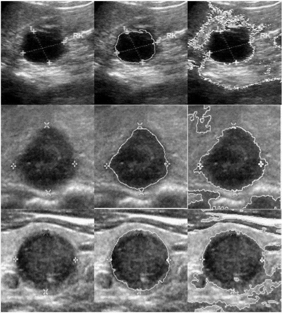
[0091] In this embodiment, a semi-automatic image segmentation method based on a point-based distance function shape constraint is applied to the segmentation of ultrasound images of thyroid nodules, such as figure 2 As shown, the first column is the original ultrasound image, the second column is the corresponding segmentation result map in this embodiment, and the last column is the segmentation result map using the method in literature [1]. Since the energy functional in the literature [1] has no shape constraint term, all the obtained results are over-segmented.

Embodiment 2

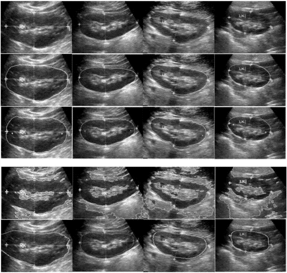
[0093]In this embodiment, the semi-automatic image segmentation method based on the shape constraint of the distance function of two points is applied to the segmentation of the ultrasound image of the kidney.

[0094] Such as image 3 As shown, the first row of images is the original kidney ultrasound image, the second row is the results of manual segmentation by doctors, and the third row is the corresponding segmentation results using the two-point distance shape constraint model in the embodiment. A normal human kidney is shaped like a pea. The shape generated by the point distance function based on two points of the present invention is a series of ellipses a priori. The fourth row is a schematic diagram of the segmentation results of the algorithm in [1]. The last line is a schematic diagram of the segmentation results of the algorithm in [3]. Literature [3] introduces a parameterized hyperellipse shape prior in the variational framework to segment ultrasound kidney i...


PUM


Abstract

The invention discloses a semi-automatic medical image segmentation method based on the shape constraint of a point distance function. The method comprises the following steps of selecting to-be-processed medical image, and defining an n-point distance function in the plane of the image; merging the shape constraint of the point distance function into a variation frame to obtain an activity contour model based on the shape constraint of the point distance function; solving the activity contour model; and solving a gradient flow equation to realize the segmentation of the image. According to the invention, the shapes of a circle, a quasi-circle, an ellipse, a superellipse, a curved-edge triangle, a curved-edge quadrilateral, a heart-shaped shape and the like can be flexibly described. Meanwhile, a medical image with a missed boundary can be effectively segmented. Moreover, the establishment of target shape databases or optimized shape parameters is not required.

Description

technical field [0001] The invention belongs to the technical field of medical image processing, and in particular relates to a semi-automatic medical image segmentation method based on point distance function shape constraints. Background technique [0002] In medical images, there are often cases of missing image data caused by various reasons, which is manifested in the missing boundaries of organs, tissues, and lesions on the image. At this time, it is difficult to segment the tissue or organ contour of interest only by using the grayscale information of the image. For image segmentation where the target image data is lost, a common existing segmentation method is to combine the prior shape information of the image. [0003] There are two main ways to express the prior shape in the existing image segmentation: [0004] One is to establish a database of target shapes and use data analysis (such as clustering, machine learning, etc.) to obtain the representation of the p...

Claims


Application Information

Patent Timeline
IPC IPC(8): G06T7/149
CPCG06T7/149G06T2207/10081G06T2207/30056G06T2207/30096
Inventor 刘海蓉李旭杨孝平向妮
Owner NANJING FORESTRY UNIV
Why Eureka
Try Eureka