Fluorescence in-situ hybridization detection method for lncRNA in lung cancer tissue

A technique of fluorescence in situ hybridization and detection method, which is applied in the field of molecular biology to achieve the effects of accurate detection results, simple operation and avoiding interference

Inactive Publication Date: 2017-12-15
ZHONGSHAN HOSPITAL FUDAN UNIV
View PDF3 Cites 2 Cited by
  • Summary
  • Abstract
  • Description
  • Claims
  • Application Information

AI Technical Summary

Problems solved by technology

However, in the prior art, there is no report about the lung cancer tissue lncRNA fluorescence in situ hybridization detection method of the present invention

Method used


Image

  • Fluorescence in-situ hybridization detection method for lncRNA in lung cancer tissue

Examples


Embodiment 1

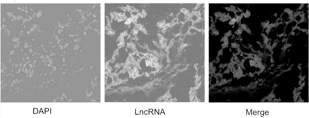
[0028] Example 1: Detection method of lncRNA fluorescence in situ hybridization in lung cancer tissue

[0029] In this embodiment, the lung cancer tissue is taken as an example to test, and the specific steps are:

[0030] 1. Preparation of frozen lung cancer tissue sections:

[0031] Fresh lung cancer tissues were sliced ​​with a frozen microtome with a thickness of 8 μm (operating temperature -20°C).

[0032] 2. Tissue fixation, digestion and permeabilization:

[0033] a. Fix with 4% paraformaldehyde at 37°C for 10 minutes;

[0034] b. Wash 3 times with 1×PBS, 3 minutes each time;

[0035] c. Collagenase IV (sigma company, product number: C5138) was incubated at 37°C for 0.3min;

[0036] d. Wash 3 times with 1×PBS, 3 minutes each time;

[0037] e. At 4°C, add 100 μl 0.5% TritonX-100 and incubate for 3 minutes;

[0038] f. Wash 3 times with 1×PBS, 3 minutes each time.

[0039] 3. LncRNA hybridization for detection:

[0040] a. Add 100 μl of pre-hybridization solution ...


PUM


Abstract

The invention provides a fluorescence in-situ hybridization detection method for lncRNA in lung cancer tissue. The method comprises the steps of preparation of a lung cancer frozen section, tissue fixation, digestion and permeation, probe hybridization detection of lncRNA, DNA staining, section sealing and fluorescence detection imaging. Expression conditions of lncRNA in the lung cancer tissue can be detected accurately and directly, and subcellular localization of lncRNA in the lung cancer tissue is shown in situ. The fluorescence in-situ hybridization detection method for lncRNA in the lung cancer tissue is simple to operate and easy to popularize and has important medical application value.

Description

technical field [0001] The invention relates to the technical field of molecular biology, in particular to a lncRNA fluorescence in situ hybridization detection method of lung cancer tissue. Background technique [0002] Lung cancer is the most common malignant tumor in my country. Studies have shown that lncRNA is involved in the occurrence and development of human malignant tumors. The role of lncRNA in lung cancer is a hotspot and difficulty in medical research. How to detect the expression of target lncRNA in lung cancer tissue is the primary problem faced by scholars. [0003] The common method for quantitative analysis of lncRNA in lung cancer tissue is RT-PCR; nucleoplasmic separation RT-PCR is used for lncRNA localization analysis. However, these experimental steps are cumbersome and complicated to operate. More importantly, the RNA extracted from lung cancer tissue often includes the RNA of tumor stromal cells, which cannot fully reflect the expression of lncRNA...

Claims


Application Information

Patent Timeline
Patent Type & Authority Applications(China)
IPC IPC(8): G01N21/64
CPCG01N21/6428
Inventor 王帅王群蒋伟时雨杨晓冬詹成蒋家好刘红汪灏姚光宇
Owner ZHONGSHAN HOSPITAL FUDAN UNIV
Why Eureka
Try Eureka