Active electrode detection device for electroretinogram and electro-oculogram

An electroretinogram and active electrode technology, which is applied in the fields of eye testing equipment, diagnostic recording/measurement, medical science, etc., can solve the problems of easy infection, poor accuracy and reproducibility, and ineffective detection of weak signals, etc., to achieve Effects of reducing wire length, improving accuracy, and eliminating noise

Active Publication Date: 2018-01-16
HUNAN MEGMEET ELECTRICAL TECH CO LTD
View PDF9 Cites 6 Cited by
  • Summary
  • Abstract
  • Description
  • Claims
  • Application Information

AI Technical Summary

Problems solved by technology

[0007] The invention solves the problems that the existing technology cannot effectively detect weak signals, and the accuracy and reproducibility are poor
On the other hand, the present invention solves the problem that electrodes are easily infected after repeated use

Method used


Image

  • Active electrode detection device for electroretinogram and electro-oculogram
  • Active electrode detection device for electroretinogram and electro-oculogram
  • Active electrode detection device for electroretinogram and electro-oculogram

Examples


Embodiment Construction

[0030] In order to make the technical means realized by the present invention clear, the present invention will be further described below in conjunction with the accompanying drawings.

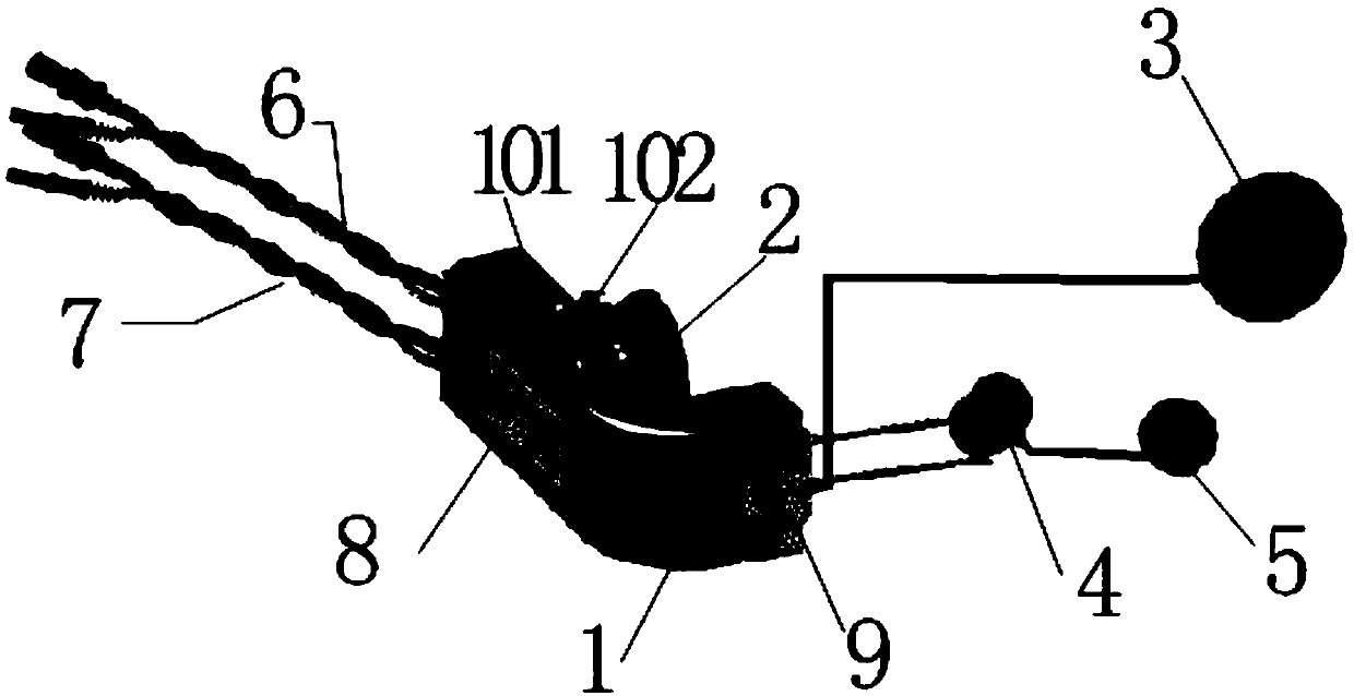
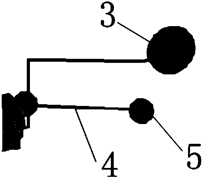
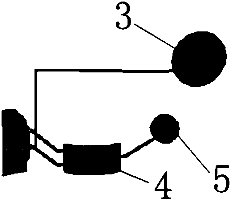
[0031] Such as figure 1 As shown, the present invention is a portable detection device for electroretinogram and electrooculogram, comprising miniature pre-signal amplifier 1, ground electrode 3, ERG electrode 4, EOG electrode 5 and reference electrode 2, said ground electrode 3, ERG electrode 4, EOG electrode 5 and reference electrode 2 are connected to the miniature pre-signal amplifier 1. ERG records the potential change of cells in the retina through the contact of the electrodes with the cornea, and the EOG contacts the skin on both sides of the orbit through the electrodes. The ERG electrode 4 and the EOG electrode 5 share a reference electrode 2 to record the change of the resting potential of the eyeball.

[0032] The ERG electrode 4 can be designed to be in contact with the eyeball ...


PUM


Abstract

The invention provides an active electrode detection device for an electroretinogram and an electro-oculogram. The device comprises a miniature front signal amplifier, a ground electrode, an ERG electrode, an EOG electrode and a reference electrode, the ground electrode, the ERG electrode, the EOG electrode and the reference electrode are connected with the miniature front signal amplifier, the reference electrode is arranged on one side of the miniature front signal amplifier, the ERG electrode and the EOG electrode use the same universal reference electrode, and the reference electrode is attached to the surface of human face skin. According to the device, a short connecting wire between a sensor and the amplifier is provided with a short transmission line, so that most external noise and interference can be eliminated; by means of the design, signals can have high signal-to-noise ratio, high reproducibility, high common-mode rejection ratio and high sensibility to low stimulation, and the electrodes can be made into disposable electrodes, so that infection is prevented, which is beneficial to patients and doctors, and infection control standards of a hospital are met.

Description

[technical field] [0001] The invention relates to the technical field of ophthalmic medical treatment, in particular to an active electrode detection device for electroretinogram and electrooculogram with a preamplifier. [Background technique] [0002] Electroretinogram (ERG) has a history of 100 years. At present, it is widely used in clinical ophthalmology. It records the changes of retinal potential through the surface of the eyeball, so as to monitor, evaluate and diagnose the health status of the retina. This technique requires the use of electrodes to record ERG. The positive electrode can be placed on the surface of the eyeball or the corresponding part using a corneal contact or non-corneal contact electrode. The reference electrode and the ground electrode can be placed on the corresponding skin, such as the earlobe and forehead. When light is stimulated, the total electrical response of the retina recorded by the positive electrode from the cornea or corresponding ...

Claims


Application Information

Patent Timeline
Patent Type & Authority Applications(China)
IPC IPC(8): A61B5/0496A61B3/10
CPCA61B3/10A61B5/398A61B5/297
Inventor 赵英军马腾·艾瑞克·柏伦
Owner HUNAN MEGMEET ELECTRICAL TECH CO LTD
Why Eureka
Try Eureka