
Endoscopic image processing method and system

An image processing and endoscopy technology, applied in the field of image processing, can solve problems such as time-consuming for doctors, difficult to accurately identify images, and no help in diagnosing diseases, so as to reduce the workload of processing, reduce the workload of doctors, and improve the efficiency of doctors Effect

Inactive Publication Date: 2018-03-06
EZHOU INST OF IND TECH HUAZHONG UNIV OF SCI & TECH +1
View PDF7 Cites 21 Cited by
  • Summary
  • Abstract
  • Description
  • Claims
  • Application Information

AI Technical Summary

Problems solved by technology

The patient swallows the capsule with water like taking medicine, and it wriggles with the gastrointestinal muscles, records the pathological images in the digestive tract through the built-in micro camera, and displays the images to the doctor to provide a basis for his diagnosis, but because the capsule endoscope Relying on intestinal peristalsis in the body, the obtained images are often blurred, which may lead to unclear images of pathological areas and no way to accurately diagnose. For the problem that capsule endoscopic images are difficult to accurately identify, there is currently no special identification method. Most of them are identified by the naked eye of doctors. With the denoising and enhancement of images, the accuracy of identification can be improved to a certain extent, but the workload is huge, and manual identification is difficult to avoid false detection and missed detection, which brings great harm to the diagnosis of diseases. to a certain degree of difficulty
In addition, the capsule endoscope relies on gastrointestinal peristalsis to take pictures of the inner wall of the human digestive tract. The working time can last for 6-8 hours, and a single inspection will produce 30,000-80,000 pictures. At certain moments, its forward speed is relatively slow, resulting in no significant difference between adjacent images, resulting in a large number of redundant images with high similarity, which are not helpful for doctors to diagnose diseases and will consume time for doctors; doctors It is difficult to find lesion pictures in a large number of pictures, and the workload is heavy

Method used


Image

  • Endoscopic image processing method and system
  • Endoscopic image processing method and system
  • Endoscopic image processing method and system

Examples


Embodiment Construction

[0020] In order to make the object, technical solution and advantages of the present invention clearer, the present invention will be further described in detail below in conjunction with the accompanying drawings and embodiments. It should be understood that the specific embodiments described here are only used to explain the present invention, not to limit the present invention.

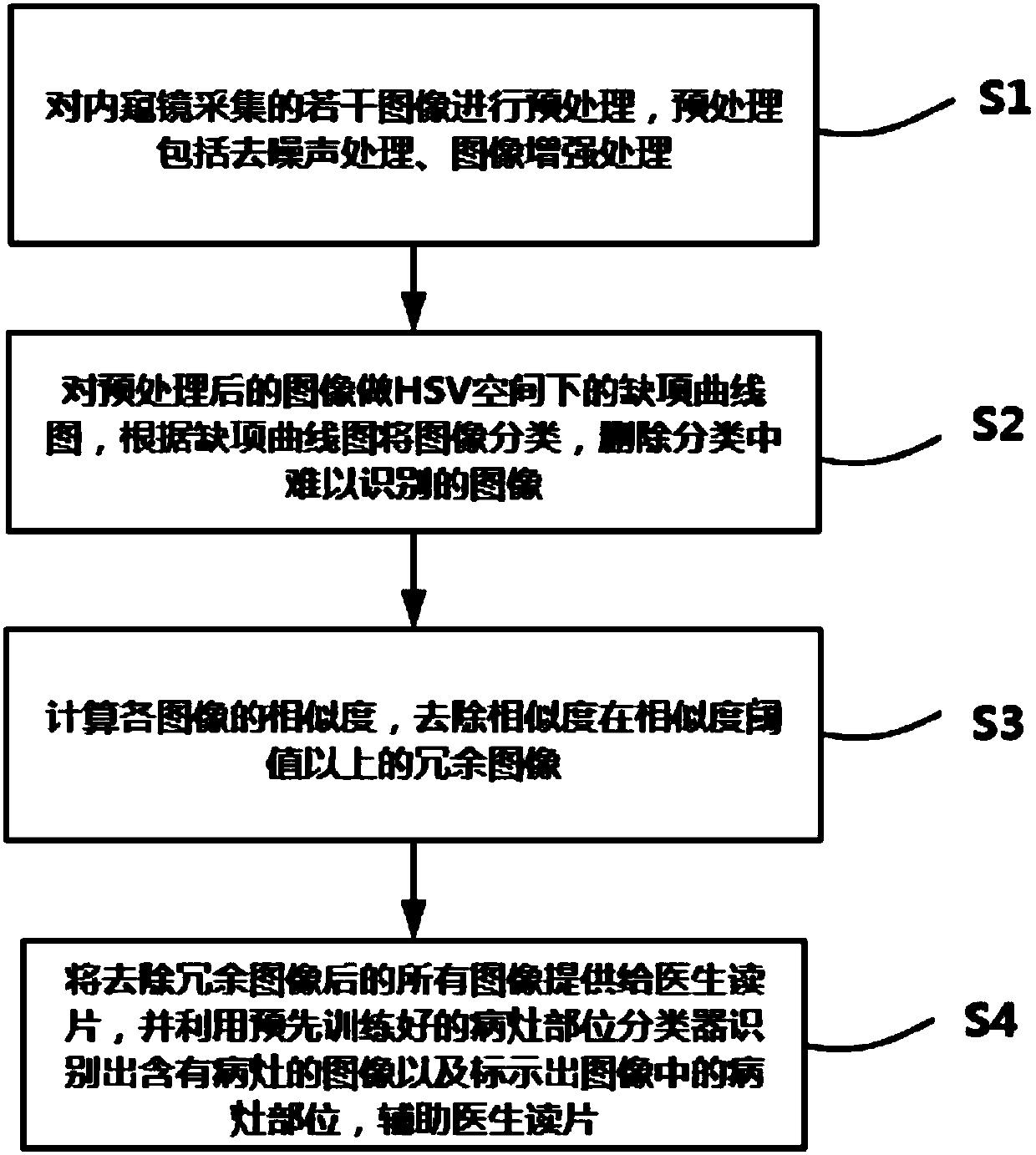
[0021] The invention provides an endoscope image processing method, comprising:

[0022] S1. Preprocessing several images collected by the endoscope, the preprocessing includes denoising processing and image enhancement processing;

[0023] S2. Perform a missing item curve under the HSV space on the preprocessed image, classify the image according to the missing item graph, and delete images that are difficult to identify in the classification;

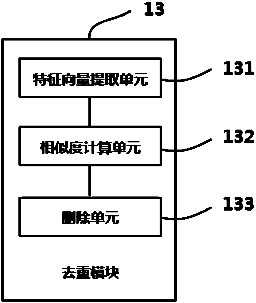
[0024] S3. Calculate the similarity of each image, and remove redundant images whose similarity is above the similarity threshold;

[0025] S4. Provide ...


PUM


Abstract

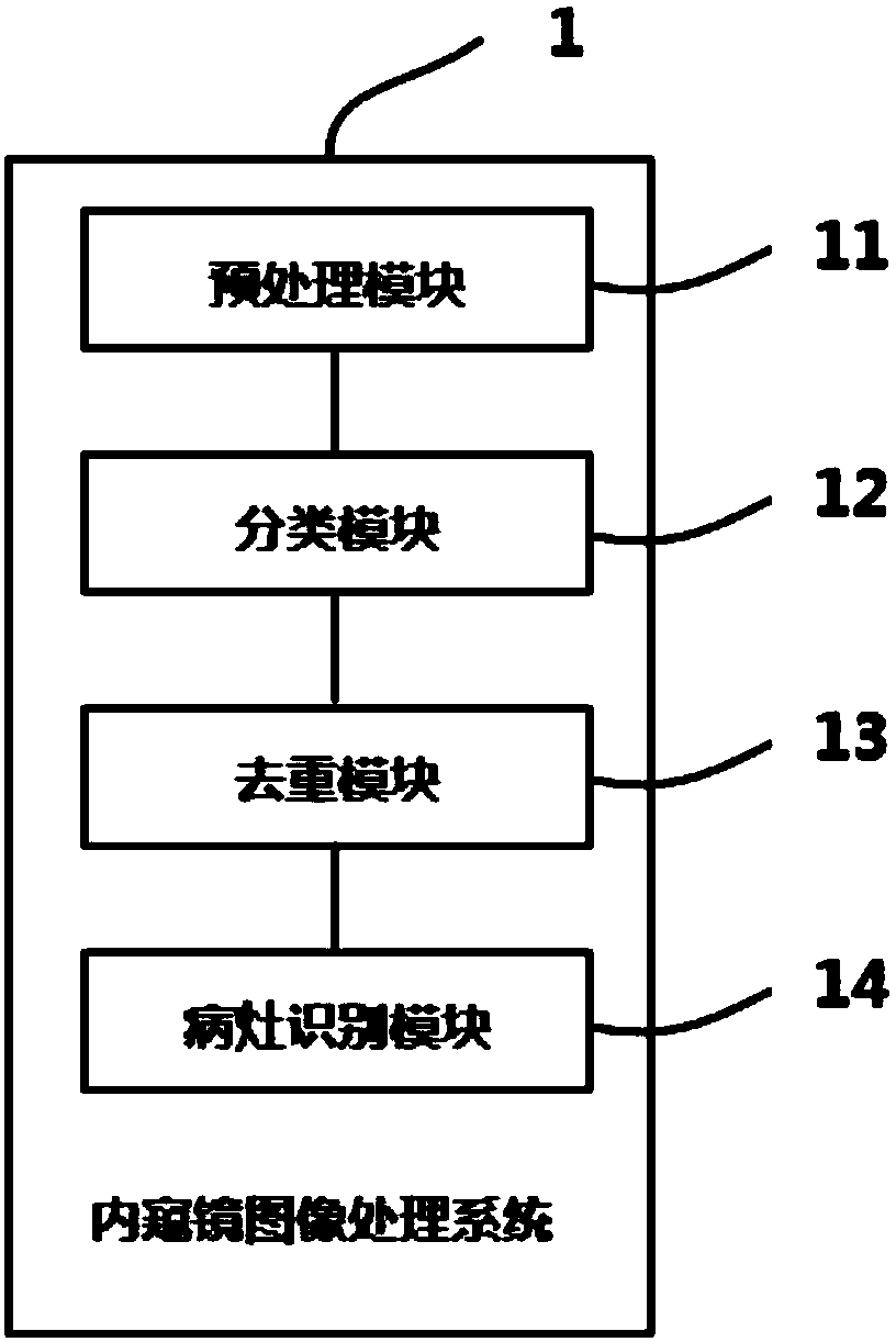
The invention discloses an endoscopic image processing method and system. The method comprises the following steps: carrying out pretreatment on a plurality of images collected by an endoscope; carrying out classification on the endoscope images obtained after pretreatment according to a missing item curve graph under an HSV space, and deleting images hard to recognize, wherein the images hard torecognize are fuzzy, and are of no effect for focus judgment, so that the images hard to recognize can be deleted to reduce follow-up image processing workload; calculating image similarity, and deleting redundancy images, the similarity of which is beyond the similarity threshold, wherein the redundancy images, the similarity of which is larger, are of no effect for focus judgment, so that by deleting the redundancy images and keeping effective images, follow-up image processing workload can be reduced, image analysis processing speed is accelerated and doctor's workload is relieved; and training a focus position classifier, and recognizing images having focus through the focus position classifier and marking focus positions in the images to assist doctors to read films. The method improves efficiency of the doctors and prevents the doctors from causing false inspection and missing inspection due to too much workload.

Description

technical field [0001] The invention relates to the technical field of image processing, in particular to an endoscope image processing method and system. Background technique [0002] Gastrointestinal mucosal disease and the resulting cancer of the digestive tract are one of the biggest killers of national health (accounting for about 60-70% of all malignant tumors in my country). According to statistics from the American Cancer Society, early detection and diagnosis of neoplasia metastasis is a key factor in reducing the mortality rate of digestive tract cancer (especially bowel cancer): If the disease is detected in the early stage and treated, the five-year survival rate can usually exceed 90%; if it is not detected in the early stage but allowed to develop to the middle and late stage, the five-year survival rate of the patient is only less than 10%. Traditionally, doctors need to insert a fiberoptic endoscope into the patient's body to observe the inside of the digest...

Claims


Application Information

Patent Timeline
IPC IPC(8): G06T7/00G06T5/00G06T7/90G06K9/62G06T7/45
CPCG06T7/0012G06T7/45G06T7/90G06T2207/10024G06T2207/10068G06T2207/30028G06T2207/30092G06F18/22G06F18/24G06T5/94
Inventor 马骁萧冯宇付玲
Owner EZHOU INST OF IND TECH HUAZHONG UNIV OF SCI & TECH
Learn More