

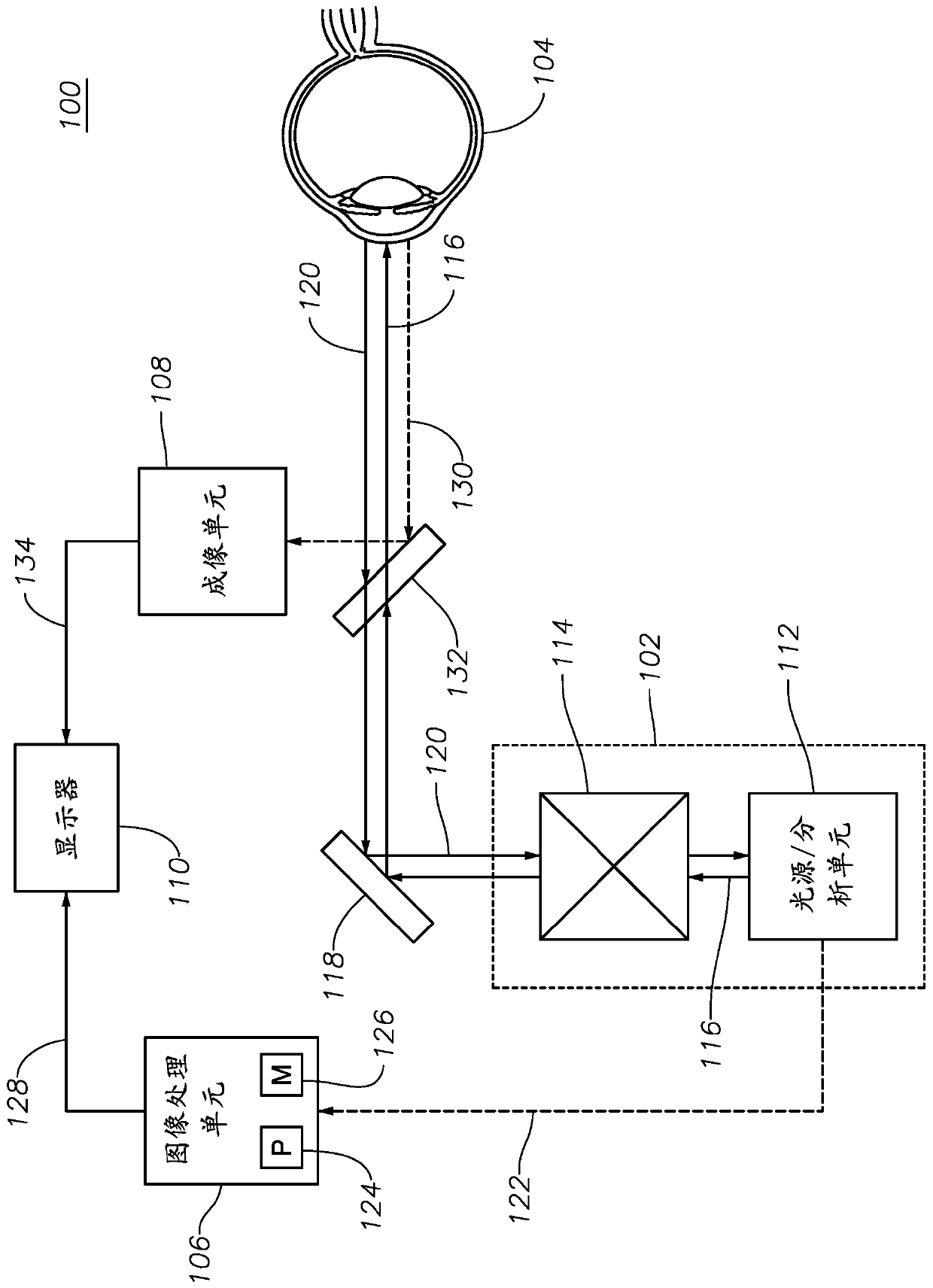
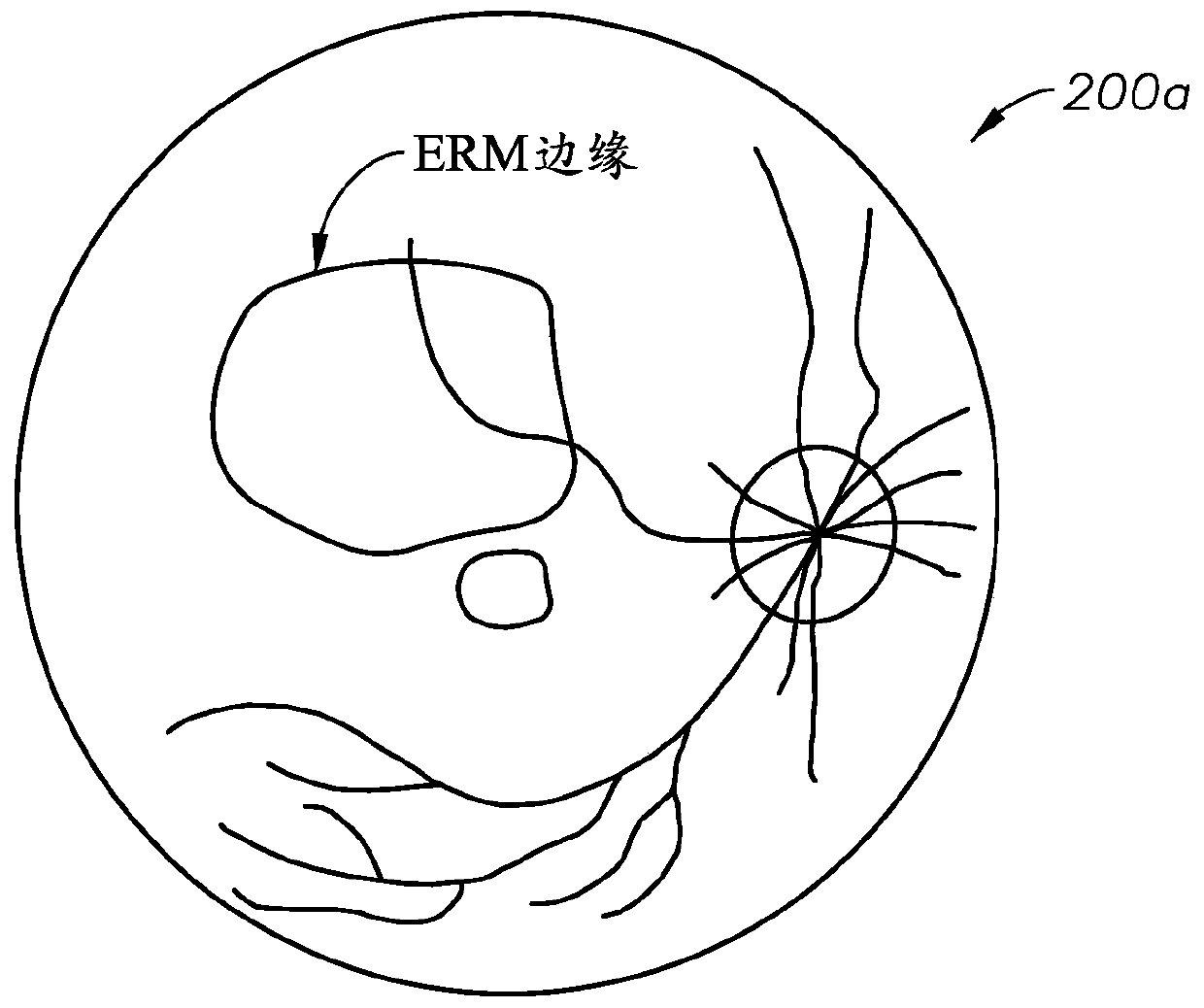
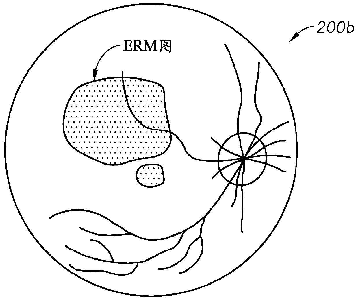
Epiretinal membrane peeling guided by optical coherence tomography
An epiretinal membrane, imaging unit technology, applied in optics, laser surgery, optical components, etc., can solve the problem of unclear potential toxicity of retinal cells, and achieve the effect of reducing risks
- Summary
- Abstract
- Description
- Claims
- Application Information
AI Technical Summary
Problems solved by technology
Method used
Image

Examples
Embodiment Construction
[0011] For the purpose of promoting an understanding of the principles of the present disclosure, reference will now be made to the embodiments shown in the drawings, and specific language will be used to describe the embodiments. However, it should be understood that it is not intended to limit the scope of this disclosure. A person of ordinary skill in the field involved in the present disclosure will generally be fully able to think of any changes and further modifications to the described systems, devices, and methods, and any further application of the principles of the present disclosure. Specifically, it is entirely conceivable that the system, device, and / or method described in one embodiment can be combined with the features, components, and / or steps described in other embodiments of the present disclosure. However, for the sake of brevity, the numerous repetitions of these combinations will not be described separately. For simplicity, in some cases, the same referenc...
PUM
 Abstract
Description
Claims
Application Information