Method for evaluating skin conditions by skin keratinocyte form
A technology of keratinocytes and chamfering, which is applied in material analysis by optical means, preparation of test samples, measurement devices, etc., and can solve problems that affect test results, easily generate bubbles, and appear unclear
- Summary
- Abstract
- Description
- Claims
- Application Information
AI Technical Summary
Problems solved by technology
Method used
Image
Examples
Embodiment 1
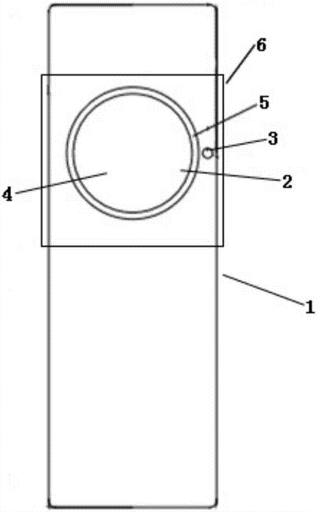
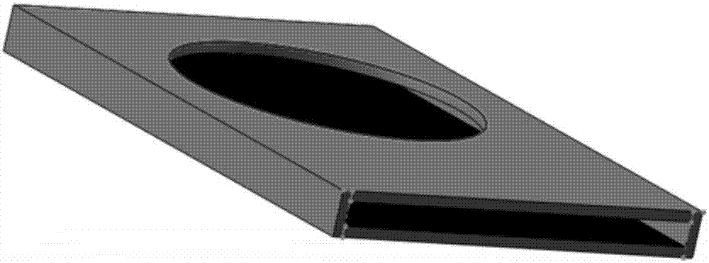
[0091] see figure 1 , a keratinocyte sampling detection tool, comprising a loading plate 1, a scotch tape 2 and a small hole 3, wherein the loading plate is provided with a collection hole 4 with a radius of 9 mm, and one side of the collection hole 4 is provided with a height of 1mm chamfer 5, the adhesive layer of the scotch tape is fixed on the non-chamfered side of the collection hole, and the scotch tape completely covers the collection hole. The loading board 1 is a PC board with a length of 76.2mm, a width of 25.48mm and a thickness of 2mm, and the scotch tape is a test film for exfoliation of skin horny layer cells from CK Company of Germany. The diameter of the small hole is 1.35 mm, and the center of the collection hole is at the same height on the loading plate.
Embodiment 2
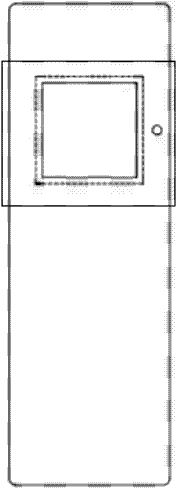
[0093] see figure 2 , a keratinocyte sampling detection tool, comprising a loading plate 1 and scotch tape 2, wherein the loading plate is provided with a collection hole 4 (square) with a length of 9 mm on one side, and one side of the collection hole 4 is provided with a height of 1mm chamfer 5, the adhesive layer of the scotch tape is fixed on the non-chamfered side of the collection hole, and the scotch tape completely covers the collection hole. The loading board 1 is a PC board with a length of 76.2mm, a width of 25.48mm and a thickness of 2mm, and the scotch tape is a test film for exfoliation of skin horny layer cells from CK Company of Germany.
Embodiment 3
[0095] Volunteer A, female, 30 years old, uses the keratinocyte sampling detection tool of the present invention to evaluate the skin condition, including the following steps:
[0096](1) Sampling: Press the chamfered side of the keratinocyte sampling detection tool in Example 1 on the skin, align the small hole with the intersection of the corner of the eye and the wing of the nose, and stroke it from top to bottom with the fingertips Press the collection hole three times; hold down around the corner of the eye with your hand, and slowly tear off the tool from bottom to top at the same time;
[0097] (2) Ferrous sulfate dyeing: the ferrous sulfate solution with a concentration of 0.01g / ml is dropped into the collection hole from the side with the chamfer, and heated by microwave at a temperature of 50°C. After 6 minutes of dyeing time, use Wash with distilled water, wash three times, three minutes each time;
[0098] (3) Counterstaining with Nuclear Fast Red: Drop Nuclear Fa...
PUM
| Property | Measurement | Unit |
|---|---|---|
| Concentration | aaaaa | aaaaa |
| Height | aaaaa | aaaaa |
| Width | aaaaa | aaaaa |
Abstract
Description
Claims
Application Information
 - R&D
- Intellectual Property
- Life Sciences
- Materials
- Tech Scout
- Unparalleled Data Quality
- Higher Quality Content
- 60% Fewer Hallucinations
Browse by: Latest US Patents, China's latest patents, Technical Efficacy Thesaurus, Application Domain, Technology Topic, Popular Technical Reports.
© 2025 PatSnap. All rights reserved.Legal|Privacy policy|Modern Slavery Act Transparency Statement|Sitemap|About US| Contact US: help@patsnap.com


