Retractable brush for use with endoscope for brush biopsy

a brush biopsy and endoscope technology, applied in medical science, surgery, vaccination/ovulation diagnostics, etc., can solve the problems of ineffective treatment of patients, difficult determination of esophagus cancer, and inability to improve the mortality rate of oral cancer in the last 20 years

Inactive Publication Date: 2006-02-28
GOLDSTEIN FAMILY PARTNERSHIP L P AS AGENT FOR THE SECURED PARTIES +2
View PDF2 Cites 42 Cited by
  • Summary
  • Abstract
  • Description
  • Claims
  • Application Information

AI Technical Summary

Benefits of technology

[0042]The structure of the brush and bristles including the stiffness thereof as well as the shape of the bristle tips contribute to the effectiveness of the brushing or scrubbing action in retrieving cells from the transepithelial layers. The shape of the bristle tips is determined by the bristle cutting process. The bristle tips, preferably, have scraping edges. The tips of the brush and the brush itself may be considered as an assemblage of penetrating edges.

Problems solved by technology

Cancers of the esophagus are also difficult to determine and are frequently not observable until an advanced state of the disease, often being too late for the patient to be effectively treated.
Despite advances in surgery, radiation, and chemotherapy, the mortality rate of oral cancer has not improved in the last 20 years.
There are several reasons for the high mortality rate from oral cancer, but undoubtedly, the most significant factor is delayed diagnosis.
Unfortunately, approximately two thirds of patients at time of diagnosis have advanced disease, and over 50% display evidence of spread to regional lymph nodes and distant metastases.
Delay in the diagnosis of oral and pharynx cancer is often the result of the limited diagnostic tools available in the prior art.
The dentist or physician who detects such a lesion which is not clearly suggestive of a precancer or cancer clinically, and who is limited to the prior art tools and methods, is faced with a quandary.
The overwhelming majority of these lesions are relatively harmless; however, the multitude of poorly defined lesions in the oral cavity can be confounding to the clinician.
Benign tumors, reactive processes, traumatic lesions, oral manifestations or systemic diseases, inflammatory oral disorders, and bacterial, viral and fungal infections all display similar oral features thereby impeding establishment of an accurate clinical diagnosis.
This prior art form of oral surgical biopsy is generally performed by a surgeon, and is often inconvenient, painful, and expensive.
Since the majority of oral abnormalities detected clinically prove benign when tested microscopically, and given the limitations of biopsy, including cost, inconvenience, pain and potential for complications, relatively few oral lesions are subjected to biopsy.
As a result, otolaryngology currently suffers from the effects of the same diagnostic dilemma which affects dentistry, i.e. the inability to clinically distinguish between common benign-appearing lesions and identically appearing pre-cancerous and early cancerous lesions.
In contrast, a scalpel biopsy in many areas of the throat cannot be performed as an office procedure.
This is because a scalpel biopsy in many areas of the throat may result in potentially dangerous aspiration of blood if the procedure is not performed under general anesthesia.
Referral of the patient for an operating room procedure requiring general anesthesia is both expensive and intrusive, and may expose the patient to other risks such as anesthesia and infection risk.
Abrasion of the underlying cervical epithelium is undesired, as it can cause bleeding and discomfort to the patient.
Although cytology has been adopted for use in several other body sites, it has not been found useful to test questionable lesions of the oral areas.
This is in large part due to the fact that the prior art devices and methods used to obtain a cellular sample for cytology are unsatisfactory when used to sample lesions of the oral and nasal areas and areas containing similar epithelia.
While abnormal cells can spontaneously exfoliate to the epithelial surface and be gently removed by prior art instruments in the uterine cervix and other similar tissues, in many oral cavity lesions the abnormal cells never reach the surface because they are blocked by the keratin layer.
This limitation is a major cause of the high false negative rate of prior art cytological testing to detect lesions of the oral cavity.
Since it is well known that dangerous, truly cancerous oral lesions may commonly be reported as “negative” using prior art cytologic sampling techniques, prior art cytologic techniques offer little as a reliable diagnostic alternative to the lacerating biopsy and histology.

Method used


Image

  • Retractable brush for use with endoscope for brush biopsy
  • Retractable brush for use with endoscope for brush biopsy
  • Retractable brush for use with endoscope for brush biopsy

Examples


Embodiment Construction

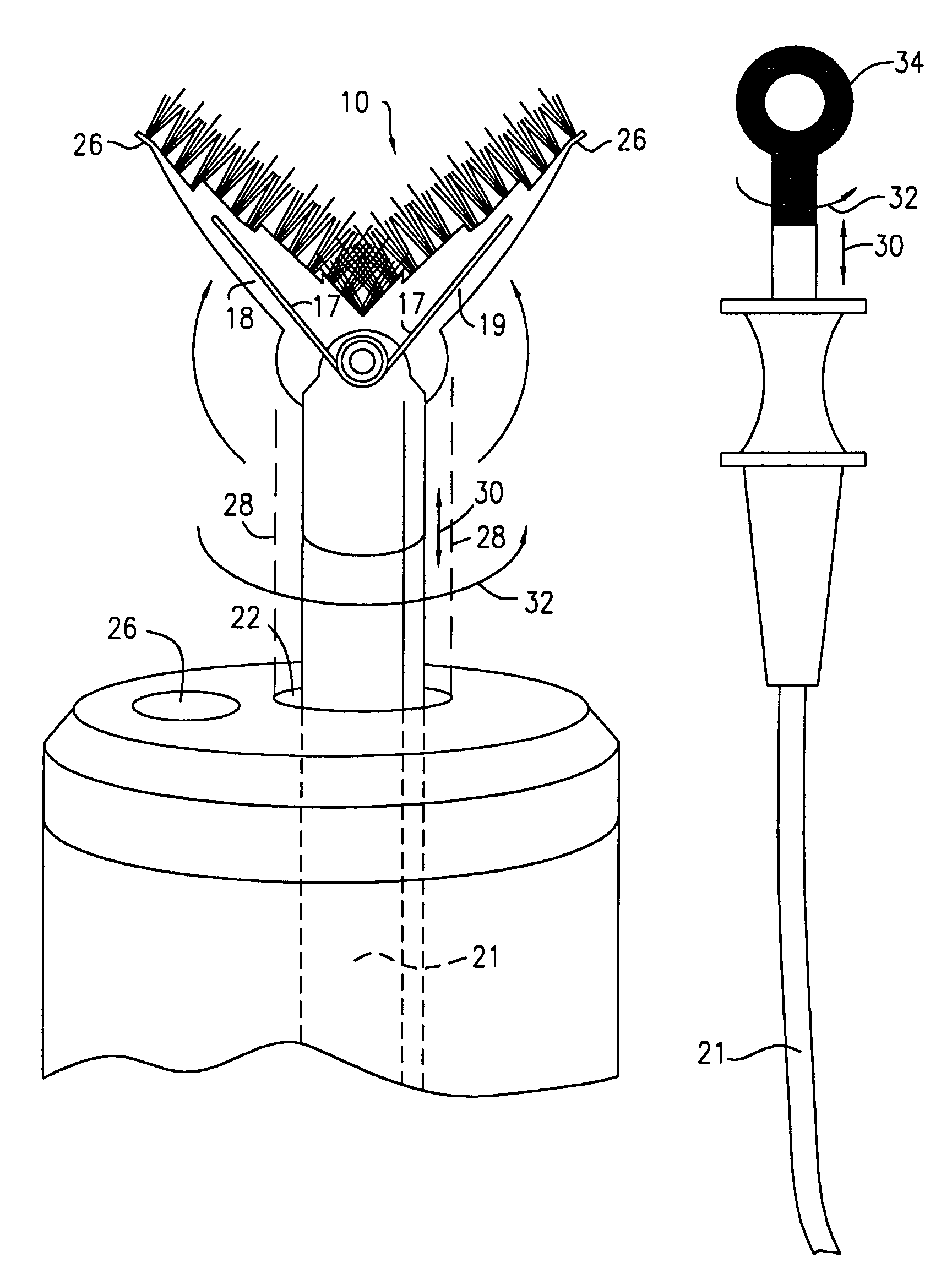
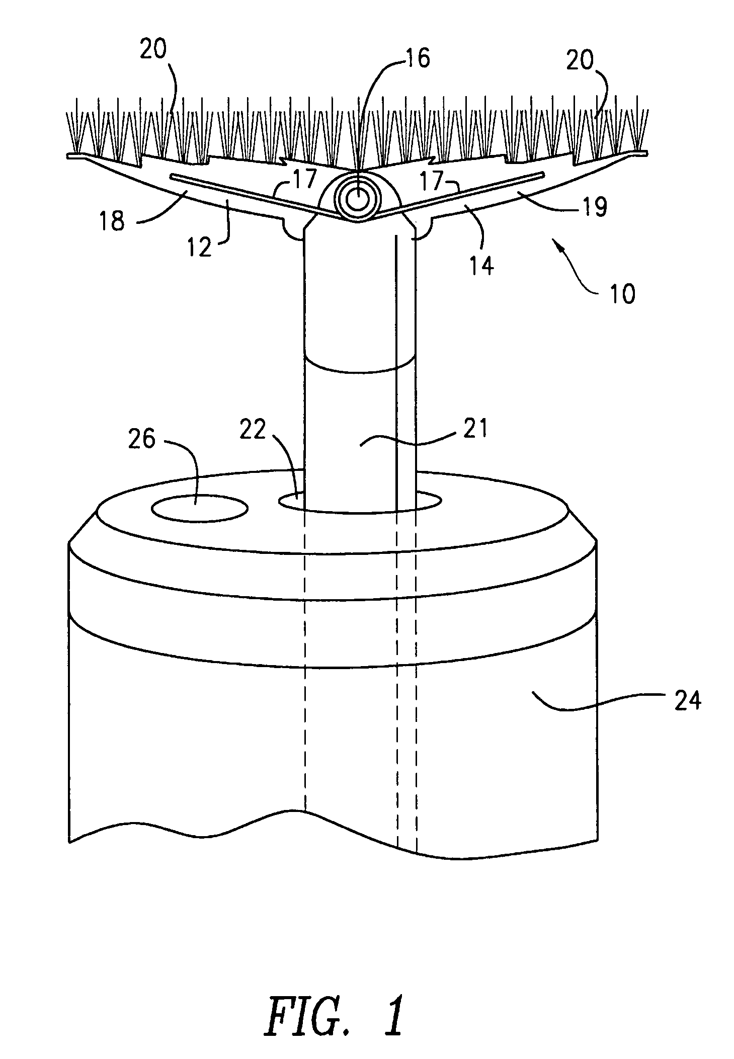
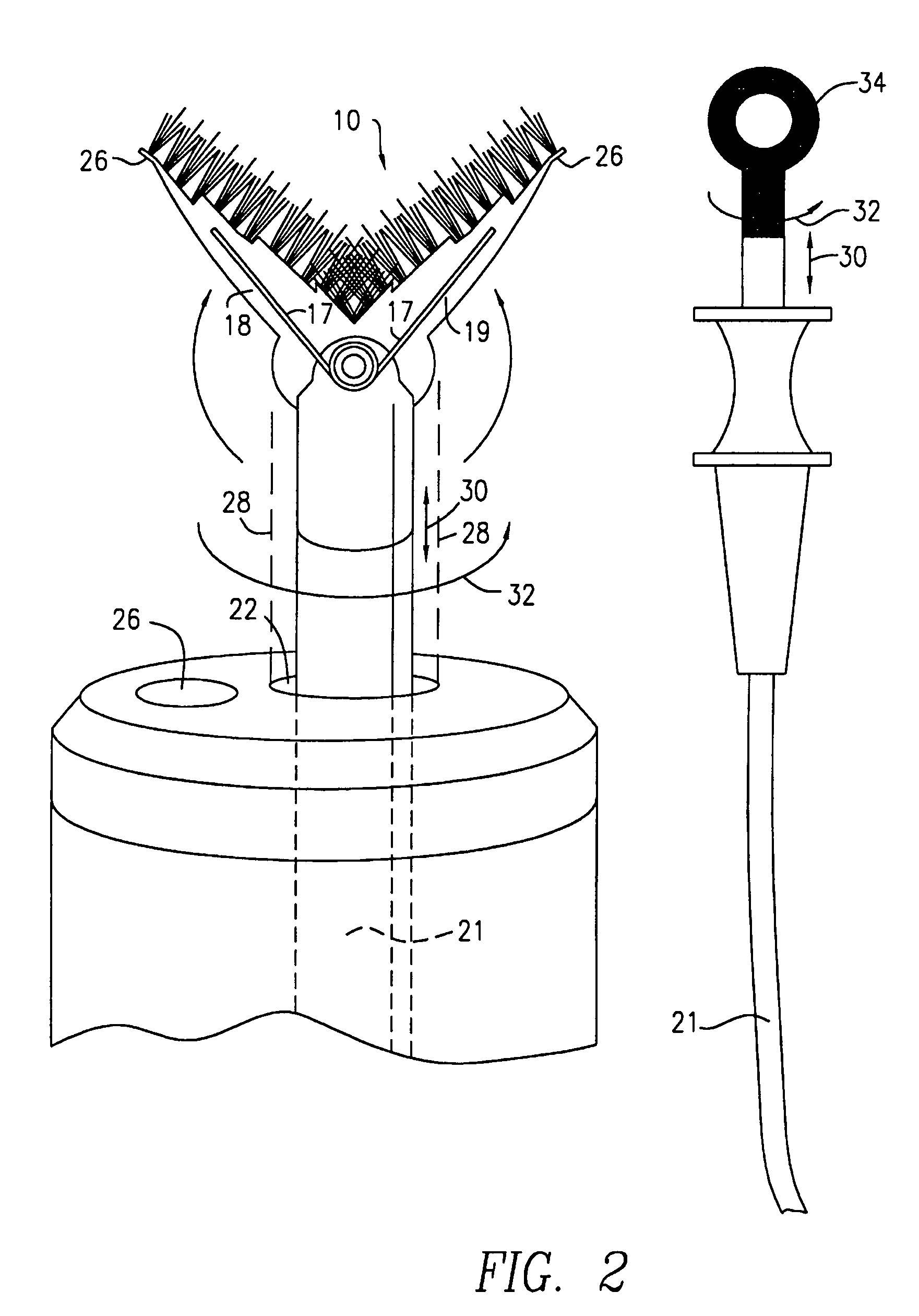
[0049]The prior art has used brushes with endoscopes to sample the examined area by moving the bristles of the brush across the suspect tissue area. Such brushes are similar to those described above which remove cells from the superficial layer, and the bristles do not penetrate below such layer because there is no direct force applied pushing tips of the bristles into the tissue being examined. Further, the prior art brushes merely rub against the surface and the brush area is very limited. The present retractable brush opens to a large size, the bristles thereof directly bear on said tissue transversely thereto and are urged further into said tissue by direct pressure. The brush is then rotated to sample a large area while concurrently reaching through the basement membrane to the basal cell layer.

[0050]FIG. 1 is a partial perspective and sectional view of an embodiment of this invention comprising a brush 10 formed of two separate brush sections 12 and 14 hingedly connected toget...


PUM


Abstract

A retractable brush structure is attached to a cylindrical rigid rod which in the closed position passes through a channel in an endoscope. After the brush passes through the endoscope, the brush moves against the tissue in order to remove cells from an area under examination. The brush is withdrawn from the endoscope and sample tissue is removed from said brush for examination after it is withdrawn from the endoscope.

Description

RELATED APPLICATION[0001]This is a continuation of U.S. patent application Ser. No. 10 / 321,010, filed Dec. 17, 2002, which is to issue on Jan. 13, 2004 as U.S. Pat. No. 6,676,609, which was a continuation-in-part of prior U.S. patent application Ser. No. 09 / 849,085 filed May 4, 2001, which issued as U.S. Pat. No. 6,494,845 on Dec. 17, 2002, each of which are incorporated herein by reference.FIELD OF THE INVENTION[0002]The present invention is directed to a method and apparatus for obtaining transepithelial specimens of body surfaces using a non-lacerating technique. Specifically, the invention is directed to retractable tools such as a brush, used with endoscopes, for sampling epithelium from lesions found from the nose to the throat and in similar body tissues. The invention is also directed to an improved apparatus for non-lacerational testing of lesions that involve the epithelium of the nasopharynx, hypopharynx, pharynx, trachea, larynx, and the upper esophagus.[0003]In metaplas...

Claims


Application Information

Patent Timeline
Patent Type & Authority Patents(United States)
IPC IPC(8): A61B10/00
CPCA61B10/04A61B2010/0216A61B10/06
Inventor RUTENBERG, MARKFRIST, STEPHEN
Owner GOLDSTEIN FAMILY PARTNERSHIP L P AS AGENT FOR THE SECURED PARTIES
Why Eureka
Try Eureka