Ultrasonic three-dimensional fetal imaging method and system

An imaging method and fetal technology, applied in ultrasound/sonic/infrasonic Permian technology, ultrasound/sonic/infrasonic image/data processing, ultrasound/sonic/infrasonic diagnosis, etc. In order to improve the detection rate, improve work efficiency, and reduce interactive operations, it can solve the problems of fetal area cutting and time-consuming

Active Publication Date: 2018-05-15
VINNO TECH (SUZHOU) CO LTD
View PDF5 Cites 4 Cited by
  • Summary
  • Abstract
  • Description
  • Claims
  • Application Information

AI Technical Summary

Problems solved by technology

In the traditional three-dimensional ultrasound fetal imaging system, clinicians need to interact with the interface to select ROI rectangular frames and curved frames to exclude embryos, uterine walls and other tissues; this method is time-consuming, and the ROI rectangular frame is only visible on the current scanning surface. Excluding non-fetal tissue, no guarantee that the fetus will be cropped out in other sections
[0004] Furthermore, Chinese Patent Publication No. CN 105976394 A, title: A cropping method for adaptively adjusting fetal three-dimensional ultrasound data, proposes an automatic cropping method, which first uses the graph cut theory to perform image segmentation to extract the amniotic fluid region, Then use the minimum cut method to search for the best path through the amniotic fluid region between two points, which separates the fetus; however, the success rate of this method is closely related to the image segmentation effect, and only realizes the fetal region cropping replacement of a single slice Without the manual interaction of the curve ROI, the clipping of the fetal region in the entire 3D ultrasound data cannot be done

Method used


Image

  • Ultrasonic three-dimensional fetal imaging method and system
  • Ultrasonic three-dimensional fetal imaging method and system
  • Ultrasonic three-dimensional fetal imaging method and system

Examples


Embodiment Construction

[0057] The present invention will be described in detail below in conjunction with specific embodiments shown in the accompanying drawings. But these embodiments do not limit the present invention, and the structural, method, or functional changes made by those skilled in the art according to these embodiments are all included in the protection scope of the present invention.

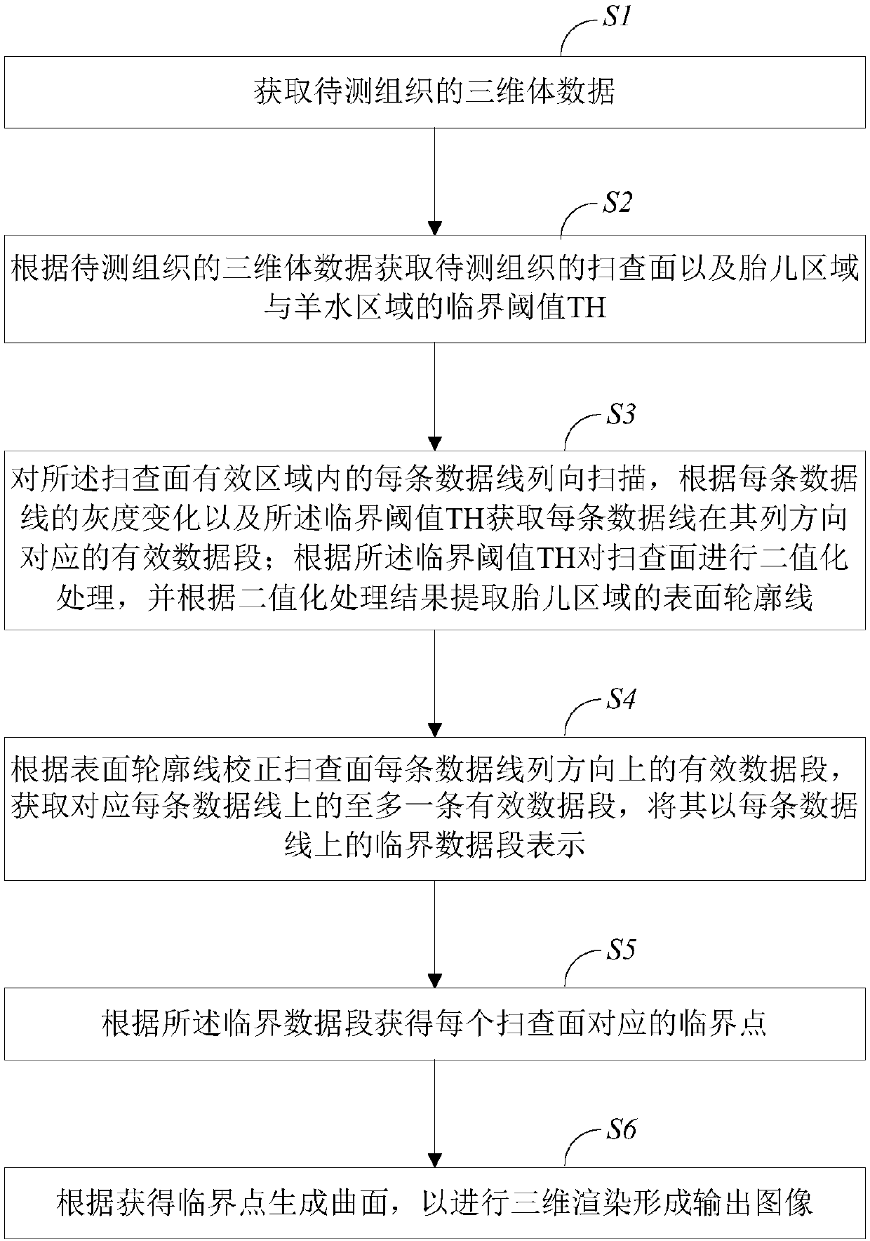
[0058] Such as figure 1 As shown, one embodiment of the present invention provides a method for ultrasonic three-dimensional fetal imaging, the method comprising:

[0059] S1. Obtain the three-dimensional volume data of the tissue to be measured, the tissue to be measured includes: the fetal region, the amniotic fluid region, and the blocking tissue region; 3D volume data.

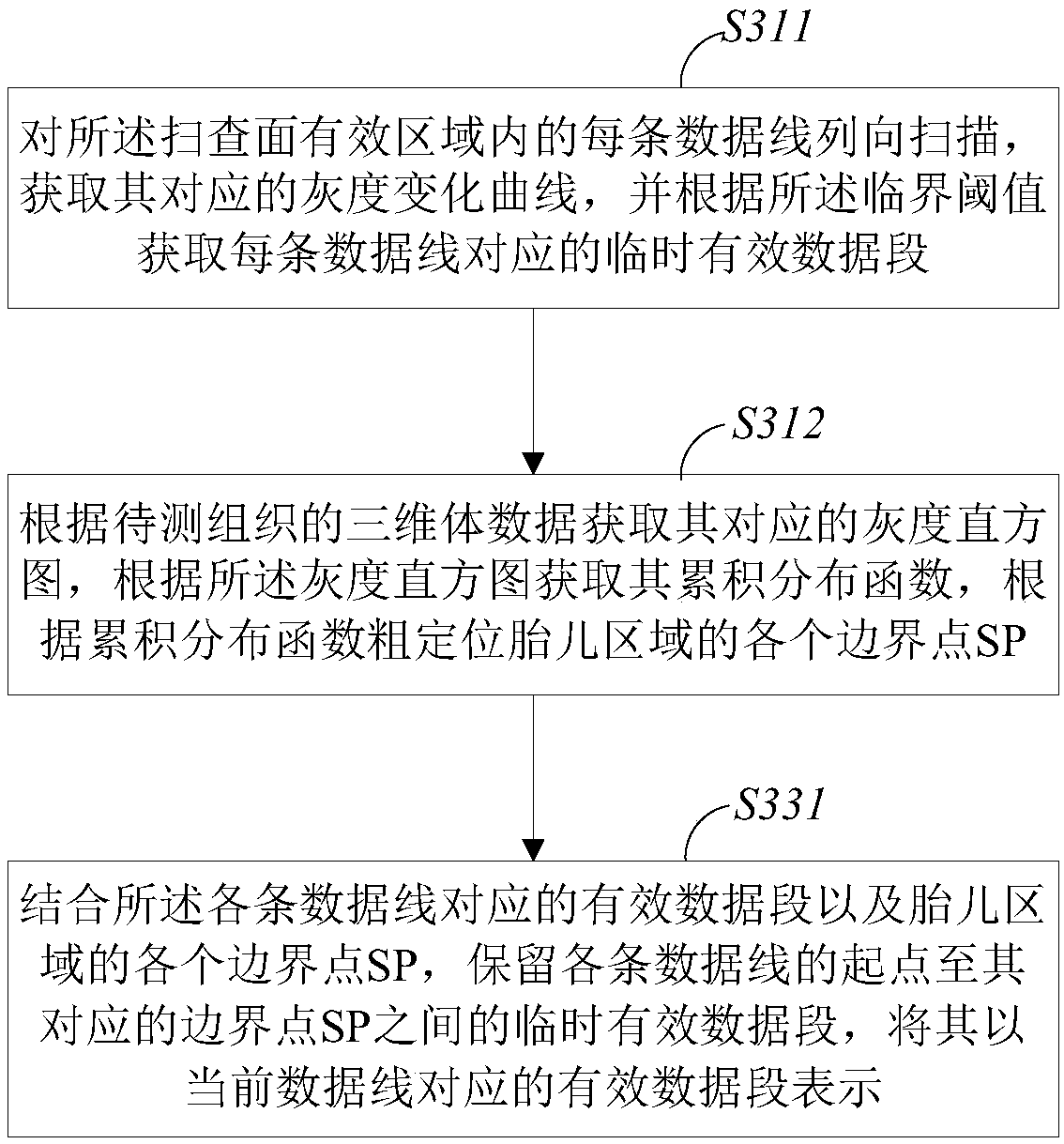
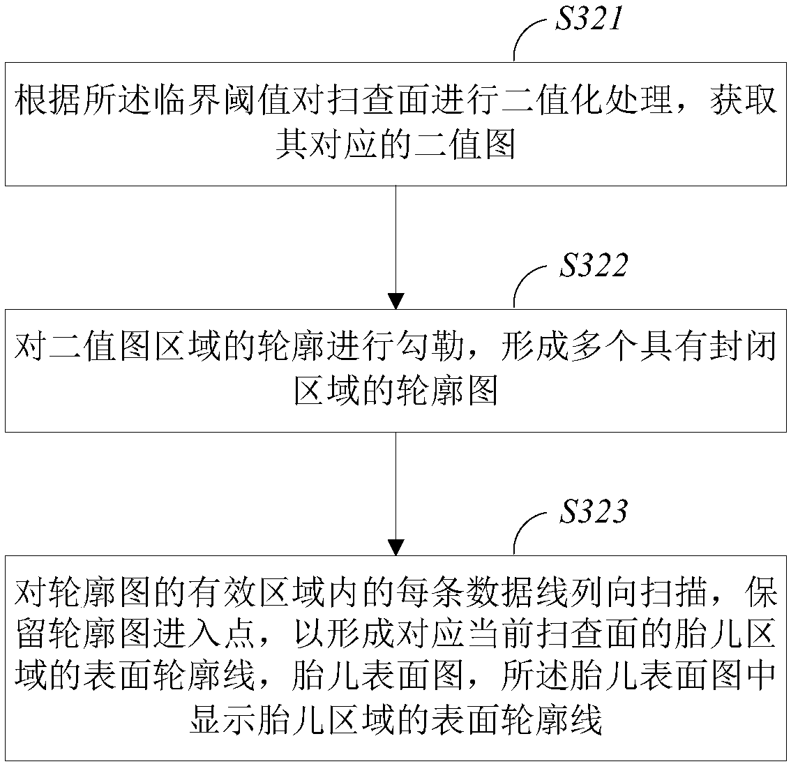
[0060] S2. Obtain the scanning plane of the tissue to be measured and the critical threshold value of the fetal area and the amniotic fluid area according to the three-dimensional volume data of the tissue to be measured, the scan...


PUM


Abstract

The invention provides an ultrasonic three-dimensional fetal imaging method and system. The method includes the steps of obtaining three-dimensional medical volume data of to-be-tested tissue, obtaining a corresponding scanning face and a critical threshold according to the volume data, scanning each data line in an effective area of the scanning face in the line direction, obtaining a corresponding effective data section according to the gray level change of each data line and the critical threshold, conducting binary treatment on the scanning face according to the critical threshold to extract a surface contour line of a fetal area, correcting the effective data section of each data line on the scanning face in the line direction according to the surface contour line, obtaining at leastone effective data section corresponding to each data line, representing the effective data section with a critical data section on each data line, obtaining a critical point corresponding to each scanning face according to the corresponding critical data section, generating a curve according to the obtained critical points, and conducting three-dimensional rendering to form an output image. The detecting rate of the fetal face and fetal structure can be improved, the interaction operation of medical staff and a machine is reduced, and work efficiency is improved.

Description

technical field [0001] The invention belongs to the field of medical ultrasonic diagnostic imaging, in particular to an ultrasonic three-dimensional fetal imaging method and system. Background technique [0002] The traditional B-mode ultrasound imaging system provides a two-dimensional image of a section of the tissue to be tested, and clinicians need to reconstruct the three-dimensional structure in the brain based on past experience; three-dimensional ultrasound imaging is usually completed based on two-dimensional images, that is, through a Combine two-dimensional images to reconstruct three-dimensional images; three-dimensional imaging can directly display the structure of the fetus or organs, making observation and clinical diagnosis more intuitive. [0003] In the process of three-dimensional tissue imaging, especially ultrasound fetal imaging, the fetal face or fetal body structure is easily blocked, which is caused by the maternal placenta, uterine wall, etc.; in or...

Claims


Application Information

Patent Timeline
Patent Type & Authority Applications(China)
IPC IPC(8): A61B8/08
CPCA61B8/0866A61B8/52
Inventor 赖昀赵一鸣
Owner VINNO TECH (SUZHOU) CO LTD
Why Eureka
Try Eureka