

Method of improving directional imaging ability of carbon dots to cell nucleolus
A technology with cell nucleolus and imaging capabilities, applied in chemical instruments and methods, measuring devices, fluorescence/phosphorescence, etc., can solve the problems of short validity period, expensive reagents, easy bleaching of fluorescence, etc., and achieve improved imaging effects and excellent directional imaging effect of effect
- Summary
- Abstract
- Description
- Claims
- Application Information
AI Technical Summary
Problems solved by technology
Method used
Image

Examples
Embodiment 1
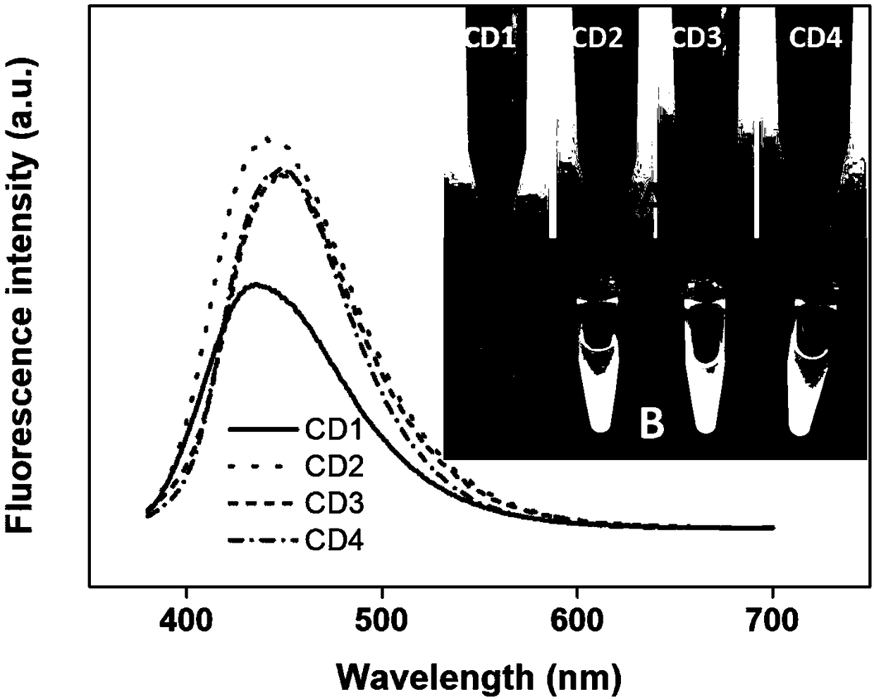
[0022] CD1 preparation:
[0023] Dissolve 1.6g of citric acid in 10mL of water, add 0.21mL of ethylenediamine to it and mix evenly to form a homogeneous solution (the molar ratio of citric acid and ethylenediamine is 1:3). The solution was transferred to a 50mL polytetrafluoroethylene reactor, and placed in an oven at 180 degrees Celsius for 12 hours. After it was cooled to room temperature, the reaction kettle was opened to obtain a black solution.
[0024] The resulting black solution was transferred to a dialysis bag with a molecular weight cut-off of 1000, and the dialysis bag was placed in a 1L beaker, with constant stirring and water changes to remove unreacted small molecules and generated ultra-small nanoparticles.
[0025] When the solution outside the dialysis bag does not deepen further 10 minutes after the water change, and the solution outside the dialysis bag has no obvious fluorescence under the ultraviolet light, it means that the dialysis is basically complet...
Embodiment 2
[0029] CD2 preparation:
[0030] Dissolve 1.6g of citric acid in 10mL of water, add 0.42mL of ethylenediamine to it and mix evenly to form a homogeneous solution (the molar ratio of citric acid and ethylenediamine is 1:2). The solution was transferred to a 50mL polytetrafluoroethylene reactor, and placed in an oven at 180 degrees Celsius for 12 hours. After it was cooled to room temperature, the reaction kettle was opened to obtain a black solution.
[0031] The resulting black solution was transferred to a dialysis bag with a molecular weight cut-off of 1000, and the dialysis bag was placed in a 1L beaker, with constant stirring and water changes to remove unreacted small molecules and generated ultra-small nanoparticles.
[0032] When the solution outside the dialysis bag does not deepen further 10 minutes after the water change, and the solution outside the dialysis bag has no obvious fluorescence under the ultraviolet light, it means that the dialysis is basically complet...
Embodiment 3
[0036] CD3 preparation:
[0037] Dissolve 1.6g of citric acid in 10mL of water, add 0.83mL of ethylenediamine to it and mix evenly to form a homogeneous solution (the molar ratio of citric acid and ethylenediamine is 2:1). The solution was transferred to a 50mL polytetrafluoroethylene reactor, and placed in an oven at 180 degrees Celsius for 12 hours. After it was cooled to room temperature, the reaction kettle was opened to obtain a black solution.
[0038] The resulting black solution was transferred to a dialysis bag with a molecular weight cut-off of 1000, and the dialysis bag was placed in a 1L beaker, with constant stirring and water changes to remove unreacted small molecules and generated ultra-small nanoparticles.
[0039] When the solution outside the dialysis bag does not deepen further 10 minutes after the water change, and the solution outside the dialysis bag has no obvious fluorescence under the ultraviolet light, it means that the dialysis is basically complet...
PUM
 Abstract
Description
Claims
Application Information