Cervicothoracic joint imaging coil component

A coil assembly and imaging technology, applied in magnetic resonance measurement, medical science, measuring devices, etc., can solve problems such as cooperative use, insufficient signal-to-noise ratio, and complex shape of the human neck, so as to achieve convenient use and storage, and improve signal quality. The effect of the noise ratio

Inactive Publication Date: 2018-06-29
北京清影华康科技有限公司
View PDF15 Cites 5 Cited by
  • Summary
  • Abstract
  • Description
  • Claims
  • Application Information

AI Technical Summary

Problems solved by technology

[0004] At present, there are no neck-thorax coils with excellent performance on the market, because the shape of the human neck is relatively complex, and there are great differences between individuals.
Most commercial head-neck-thorax integrated coils are generally too large in order to be suitable for most subjects. Although the imaging range is large, there are not many channels in the neck, and some coil units are far away from human tissue, so the signal-to-noise ratio is not enough. high
Although the commercial carotid coil improves the signal-to-noise ratio to a certain extent, the imaging range is small, and it is often difficult to cooperate with other coils.

Method used


Image

  • Cervicothoracic joint imaging coil component
  • Cervicothoracic joint imaging coil component
  • Cervicothoracic joint imaging coil component

Examples


Embodiment Construction

[0021] The specific structure and implementation of the cervical-thoracic joint imaging coil assembly of the present invention will be described in detail below with reference to the accompanying drawings.

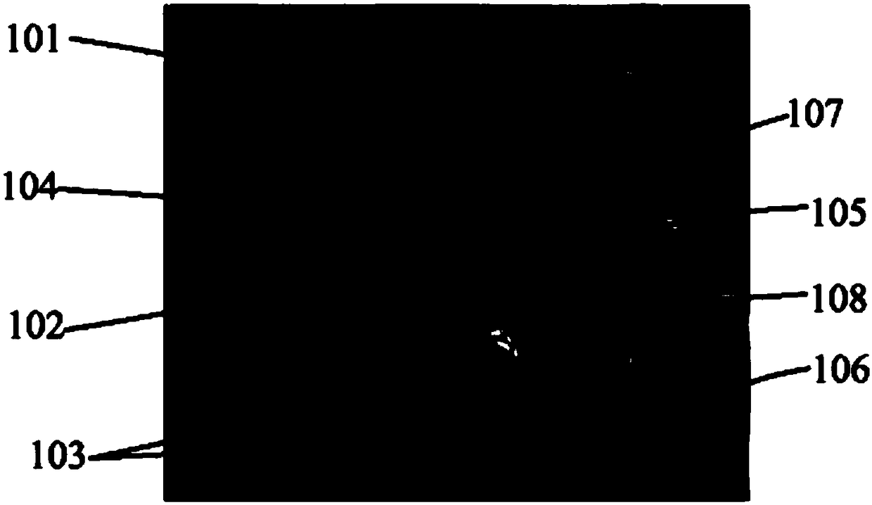
[0022] figure 1 It is the appearance structural diagram of the combined cervical and thoracic imaging coil assembly of the present invention, such as figure 1 As shown, the joint cervical and thoracic imaging coil assembly of the present invention includes a nape base part 102 placed on the bed on the back of the subject's neck and chest during imaging, and a cervical and chest front section 101 placed on the front of the subject's neck and chest. The surface of the coil is wrapped with flexible insulating material, which can make the subject feel more comfortable. The front part 101 of the neck and chest is semi-flexible, and its shape is designed according to the size of the front of the neck and chest of a standard human body. The internal skeleton support structure is...


PUM


Abstract

The invention relates to a cervicothoracic joint imaging coil component which comprises a cervicothoracic front piece, a nape base and carotid artery parts, wherein the cervicothoracic front piece isa semi-flexible structure, and the shape corresponds to the cervicothoracic front part of a standard human body; the upper surface of the nape base corresponds to the shape of the nape part of the standard human body; the lower surface of the nape base corresponds to a bed table; the carotid artery parts are movably arranged on two sides of the neck part of the nape base; coils are embedded in thecervicothoracic front piece, the nape base and the carotid artery parts; an insulating outer layer is arranged on the surface of each coil; when the cervicothoracic joint imaging coil works, the cervicothoracic front piece, the nape base and the carotid artery parts are fit with the surface of a subject.

Description

technical field [0001] The invention relates to the technical field of magnetic resonance imaging, in particular to a combined cervical and chest imaging coil assembly. Background technique [0002] The basic principle of magnetic resonance imaging is: some atoms in human tissue, such as hydrogen atoms, will generate magnetic moments due to spin motion, which can be regarded as small magnets, and these small magnets will produce directional arrangements under the action of a fixed static magnetic field And precess around the direction of the magnetic field at the same specific frequency; at this time, when a radio frequency pulse of the same frequency is applied, these hydrogen atoms absorb a certain amount of energy and generate resonance, and the spin direction is deflected under the action of the radio frequency pulse; after the radio frequency pulse disappears , these hydrogen atoms will return to their original state. During the recovery process, radio frequency energy ...

Claims


Application Information

Patent Timeline
Patent Type & Authority Applications(China)
IPC IPC(8): G01R33/34A61B5/055
CPCA61B5/055G01R33/34084
Inventor 王新元苑康
Owner 北京清影华康科技有限公司
Why Eureka
Try Eureka