
Method and system for segmenting cornea structure from OCT (Optical Coherence Tomography) cornea image

A corneal structure and image segmentation technology, applied in the field of biomedical images, can solve problems such as affecting doctors' real-time diagnosis, weak signals on both sides of the cornea, and unable to meet clinical real-time and accuracy requirements at the same time.

Active Publication Date: 2018-07-10
SHENZHEN INST OF ADVANCED TECH
View PDF4 Cites 0 Cited by
  • Summary
  • Abstract
  • Description
  • Claims
  • Application Information

AI Technical Summary

Problems solved by technology

[0004] For the low signal-to-noise ratio areas produced by anterior segment OCT, such as central corneal oversaturation artifacts, horizontal artifacts, or weak signals on both sides of the cornea due to the telecentric scanning mode (currently most frequency-domain OCT adopts this method), there is no Effective processing; the speed of existing methods is generally slow, which affects the follow-up real-time diagnosis of doctors, and cannot meet the clinical real-time and accuracy requirements at the same time

Method used


Image

  • Method and system for segmenting cornea structure from OCT (Optical Coherence Tomography) cornea image
  • Method and system for segmenting cornea structure from OCT (Optical Coherence Tomography) cornea image
  • Method and system for segmenting cornea structure from OCT (Optical Coherence Tomography) cornea image

Examples


Embodiment 1

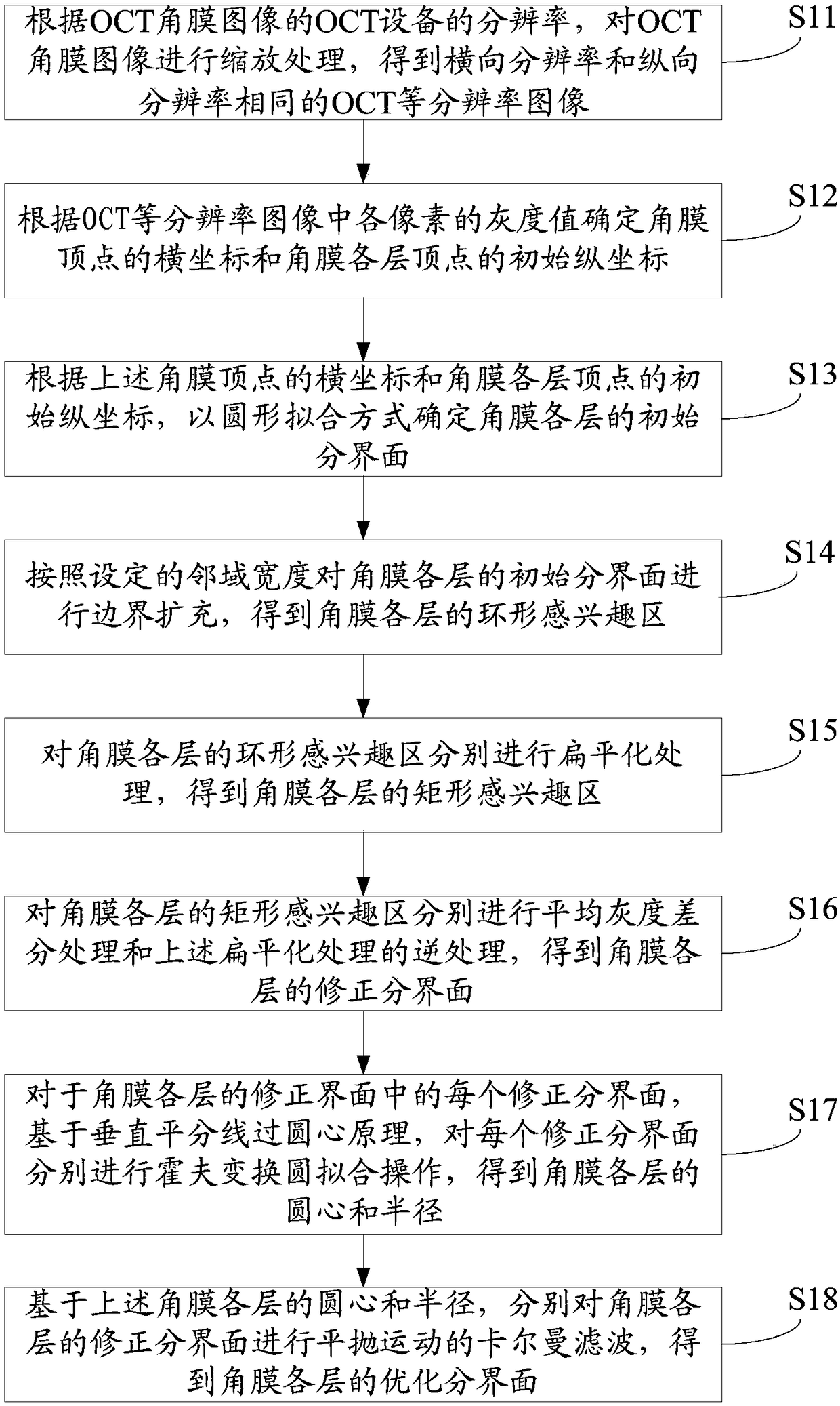
[0062] Embodiment 1 of the present invention provides a method for segmenting corneal structures from OCT corneal images, see figure 1 Shown is a schematic flow chart of a method for segmenting corneal structures from an OCT corneal image, the method comprising the following steps:

[0063] Step S11 , according to the resolution of the OCT equipment of the OCT corneal image, the OCT corneal image is scaled to obtain an OCT equal-resolution image with the same horizontal resolution and vertical resolution.

[0064] Wherein, the OCT corneal image is a two-dimensional grayscale image generated by an OCT device. Since different OCT imaging devices have different resolutions in the horizontal and axial directions, considering the subsequent circle fitting of the central cornea, the image needs to be scaled so that the image resolution is the same on the horizontal axis and the vertical axis, so that the circle in the new The image of is still a circle. The scaling process can kee...

Embodiment 2

[0163] Embodiment 2 of the present invention provides a system for segmenting corneal structures from OCT corneal images, see Figure 12 A schematic diagram of a system for segmenting corneal structures from an OCT corneal image as shown, the system includes a resolution adjustment module 121, an apex coordinate determination module 122, an initial interface determination module 123, a circular region of interest determination module 124, and a rectangular region of interest determination module 125, correction interface determination module 126, Hough transform circle fitting module 127 and Kalman filter module 128, wherein, the function of each module is as follows:

[0164] The resolution adjustment module 121 is configured to perform scaling processing on the OCT corneal image according to the resolution of the OCT device of the OCT corneal image, to obtain an OCT equal-resolution image with the same lateral resolution and longitudinal resolution. The vertex coordinate det...


PUM


Abstract

The invention provides a method and system for segmenting a cornea structure from an OCT (Optical Coherence Tomography) cornea image, and relates to the technical field of biomedical images. The method comprises the steps of scaling the OCT cornea image; determining the horizontal coordinate of the vertex of a cornea and the initial longitudinal coordinate of the vertex of each layer of the cornea; determining initial interfaces in a circle fitting mode; performing boundary expansion on the initial interface of each layer of the cornea according to the set neighborhood width to obtain annularregions of interest; obtaining rectangular regions of interest; respectively performing inverse processing of average grayscale difference processing and flattening processing on the rectangular regions of interest to obtain modified interfaces; respectively performing an Hough transform circle fitting operation on each modified interface based on a principle that the perpendicular bisector passesthe center of a circle to obtain the center and radius of each layer of the cornea; and respectively performing Kalman filtering of horizontal projectile motion on the modified interface of each layer of the cornea. The speed and accuracy of OCT cornea image for segmenting each layer of the cornea are improved according to the invention.

Description

technical field [0001] The invention relates to the technical field of biomedical images, in particular to a method and system for segmenting corneal structures from OCT corneal images. Background technique [0002] Optical coherence tomography (OCT) is a non-destructive optical signal acquisition and processing method for imaging surface tissue structure. In recent years, OCT has become an important and innovative corneal measurement and diagnostic technique. Quantitative analysis of different layers of the cornea can help diagnose changes in visual function and help doctors diagnose corneal edema, ocular hypertension, and endothelial dysfunction, as well as corneal thickness analysis before and after refractive surgery. Therefore, under the premise of ensuring speed and accuracy, it is of great significance to segment different layers of the cornea to obtain thickness and curvature information. [0003] However, the existing fully automatic corneal segmentation methods h...

Claims


Application Information

Patent Timeline
IPC IPC(8): G06T7/10G06T3/40
CPCG06T3/4023G06T2207/10101G06T2207/30041
Inventor 胡庆茂张天桥
Owner SHENZHEN INST OF ADVANCED TECH
Learn More