In vitro artificial lymph node for sensitization and expansion of t cells for therapy and epitope mapping

A technology of cells and cell groups, which is applied in the field of sensitization and expansion of T cells used in artificial lymph nodes in vitro for treatment and epitope mapping, and can solve problems such as insufficient expansion level and loss of specific function of cell death antigens

Inactive Publication Date: 2018-07-17
THE TRUSTEES OF THE UNIV OF PENNSYLVANIA
View PDF18 Cites 1 Cited by
  • Summary
  • Abstract
  • Description
  • Claims
  • Application Information

AI Technical Summary

Problems solved by technology

However, there are many technical problems in practice, including insufficient levels of expansion, premature activation-induced cell death (apoptosis), or loss of antigen specificity and / or function
Part of the problem is the inability to replicate in vitro the environment in which antigen-specific T cell expansion occurs in lymph nodes in vivo

Method used


Image

  • In vitro artificial lymph node for sensitization and expansion of t cells for therapy and epitope mapping
  • In vitro artificial lymph node for sensitization and expansion of t cells for therapy and epitope mapping
  • In vitro artificial lymph node for sensitization and expansion of t cells for therapy and epitope mapping

Examples


Embodiment 1

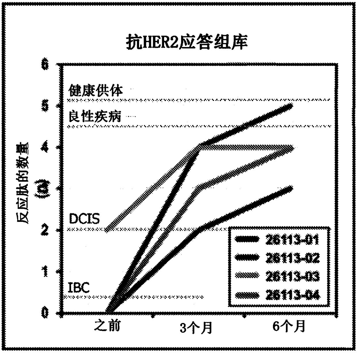
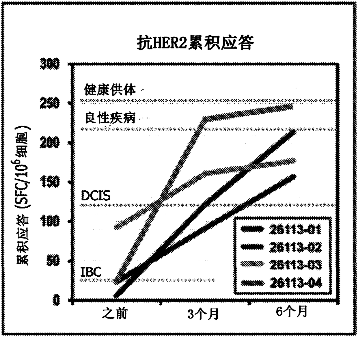
[0255] Example 1: Anti-HER2 CD4 in breast cancer patients inoculated with type I dendritic cells + Restoration of Th1 responses

[0256] The following study was designed to explore the role of helper type 1 polarized dendritic cell ("DC1") seeding.

[0257] Type I dendritic cell vaccination with HER2 with residual disease after neoadjuvant therapy + IBC patients

[0258] Four HER2 patients with residual disease after neoadjuvant therapy + IBC patients receive adjuvant HER2-pulsed type I dendritic cell vaccine. The Th1 immune response of each patient was determined before type I dendritic cell inoculation, 3 months after dendritic cell inoculation and 6 months after inoculation, and obtained from six HER2 class II peptides (SEQ ID NO: 1-6 ) pulsed patient PBMC production by measuring IFNγ production by ELISPOT as described above. Autologous type I dendritic cell vaccines were prepared as previously described. (1) overall anti-HER2 response rate (response rate >1 pepti...

Embodiment 2

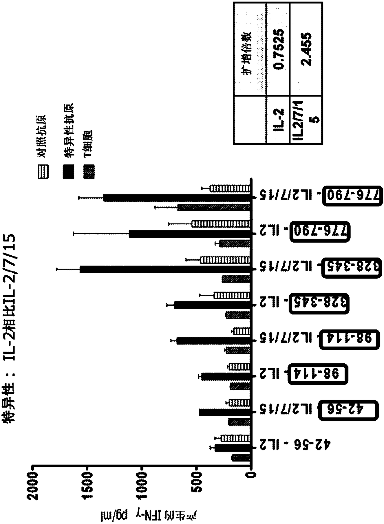
[0266] Example 2: In vitro expansion of HER2-specific Th1 cells

[0267] The following study was designed to interrogate the role of adoptive T cell transfer in restoring anti-HER2 Thl immunity. T cell expansion to levels required for adoptive therapy and epitope mapping studies while maintaining antigen specificity and cellular function. Briefly, in vitro, HER2-specific Th1 cells were generated by co-culture with HER2 peptide-pulsed type I dendritic cells and expanded using IL-2 alone or IL-2, IL-7, and IL-15 . Th1 cells were subsequently expanded by repeated HER2-peptide pulses of type I dendritic cell co-culture or by anti-CD3 / CD28 stimulation. The expansion factor was defined as: (the number of T cells after expansion / the number of T cells before expansion); the specificity of antigen-specific IIγ production was determined by ELISA.

[0268] As shown in this paper, CD4 + Repeated co-culture of T cells with HER2 peptide pulsed with type I dendritic cells stimulated wi...

Embodiment 3

[0325] Example 3: Anti-HER2 CD4 can be restored in DC1 vaccinated breast cancer patients + Th1 response

[0326] Expansion of T cell subsets is an essential step to obtain sufficient T cells for adoptive therapy or to recognize epitopes on target antigens for peptide-based vaccines. Expansion of T cells is in principle a simple process. However, in practice, there are many technical problems including insufficient levels of expansion, premature activation-induced cell death (apoptosis) or loss of antigen specificity and / or function.

[0327] Part of the problem is the inability to replicate in vitro the expansion of antigen-specific T cells that occurs in lymph nodes in vivo. These are specialized tissues that contain many different cell types besides T cells, including antigen-presenting dendritic cells, stromal cells such as epithelial cells. Each of these cell types plays a different role by providing contact-dependent (surface receptors) and soluble signals (cytokines...


PUM


Abstract

HER2+ invasive breast cancer (IBC) patients with residual disease following neoadjuvant chemotherapy have an anti-HER2 Type 1 T helper (Th1) cell immune deficit and a significant risk of recurrent disease. It has been shown that anti-HER2 CD4+ T-cell responses incrementally decrease along the breast cancer continuum - a robust response in healthy donors and patients with benign disease, a depressed response in patients with HER2+ ductal carcinoma in situ, and a nearly absent response in patients with HER2+ IBC. This invention relates to a method of creating a microenvironment for culture expansion of T cells. The expanded T cells can be used for a variety of therapeutic and research purposes.

Description

[0001] Cross References to Related Applications [0002] This application claims priority to U.S. Provisional Patent Application No. 62 / 138,684, filed March 26, 2015, and U.S. Provisional Patent Application No. 62 / 138,969, filed March 26, 2015, each of which is incorporated by reference in its entirety incorporated into this article. Background technique [0003] HER2 with residual disease + Patients with invasive breast cancer (IBC) are at significant risk for anti-HER2 type 1 T helper (Th1) cellular immunodeficiency and recurrent disease after neoadjuvant chemotherapy. Has been shown to be anti-HER2 CD4 + Statistical tapering of T cell responses along the breast cancer continuum -healthy responses in healthy donors and patients with benign disease, HER2 + Downregulated response and HER2 in patients with ductal carcinoma in situ + IBC patients are rarely responsive. [0004] The lifetime risk of developing breast cancer is almost one in eight. The erb-B2 oncogene (HER-2...

Claims


Application Information

Patent Timeline
Patent Type & Authority Applications(China)
IPC IPC(8): A61K35/17A61P35/00C12N5/074C12N15/09C12N15/85
CPCA61K35/17A61P35/00C12N5/0636C12N2501/2307C12N2501/2315C12N2502/1121C12N15/85
Inventor 布莱恩·J·赫尔尼奇莉亚·洛温菲尔德
Owner THE TRUSTEES OF THE UNIV OF PENNSYLVANIA
Why Eureka
Try Eureka