

Intelligent classification of endoscopic images and detection method of irregular lesion areas
A lesion area and irregular technology, applied in the field of medical image intelligent processing, can solve the problems of manual feature difficulty and achieve the effect of improving application value and accuracy
- Summary
- Abstract
- Description
- Claims
- Application Information
AI Technical Summary
Problems solved by technology
Method used
Image

Examples
Embodiment Construction
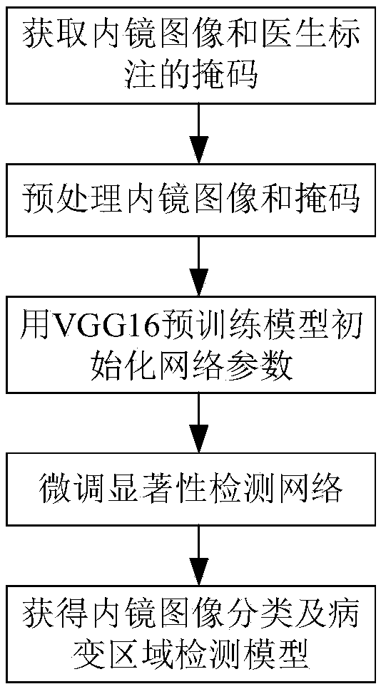
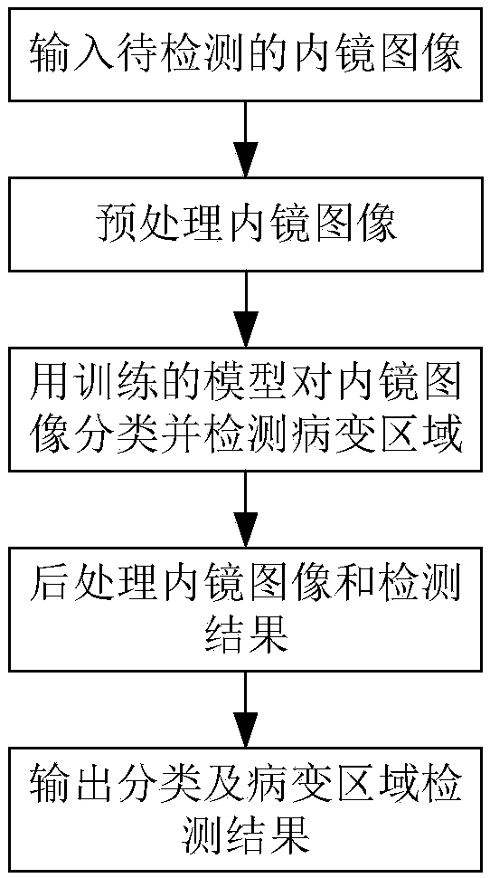
[0045] use figure 1 In the shown method, annotated endoscopic images are used to train a saliency detection network model to convergence, and the model is used for endoscopic image classification and irregular lesion region detection. For an endoscopic image to be detected, one can use figure 2 method shown. details as follows:
[0046] The specific implementation method is:
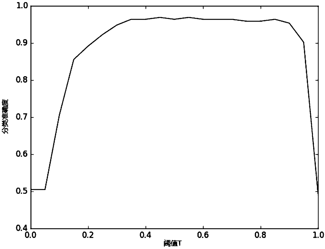
[0047] (1) Preprocess the image, according to the value of the R channel in the RGB channel of the endoscopic image (the range is 0-255), use a smaller threshold (such as 30) to binarize the image, and remove the lower value of the binarized image , and record the position information of the reserved area as (x, y, H, W);
[0048] (2) Input the preprocessed image to the network model to obtain the saliency map M, and the threshold T is set to 0.5. If there is M(i,j)>T in M, the endoscopic image is classified as lesion, otherwise it is classified as lesion. is normal; if the endoscopic image is a le...
PUM
 Abstract
Description
Claims
Application Information